
.png)
Trusted wellbutrin 300mg
Once inspection has been accomplished, examination of the bony and gentle tissue constructions follows. The area must be examined for the presence of edema, effusion, skin temperature changes, and previous websites of surgery or trauma. Systemic examination can be divided into the ankle, hindfoot, midfoot, and forefoot subgroups. Range of motion of the ankle is normally 20 degrees of dorsiflexion and forty to 50 degrees of plantarflexion. Loss of ankle dorsiflexion could also be} associated with a good Achilles tendon, posterior capsular contracture, or bony impingement. [newline]Limitation of dorsiflexion with the knee in full extension that improves passively with the knee flexed to 90 degrees signifies a contracture of the gastrocnemius muscle. Ligamentous laxity must be evaluated in comparison with the contralateral ankle joint, and palpation of the tendons must be performed to observe proof of subluxation or dislocation. Midfoot examination entails selective palpation of the bony anatomy to isolate specific joint or joint involvement. Radiology of the Foot and Ankle Radiographic research of the foot and ankle require weight-bearing X-rays when potential. The lateral view of the foot reveals the connection of the talus and calcaneus to that of the midfoot, forefoot, and ankle joint. Other research are available to assess the sesamoids, the calcaneus, or the subtalar joint. The Harris axial heel view is used to assess the calcaneal tuberosity and is important in calcaneus fractures or tarsal coalitions. Normal anatomy seen on computerized tomography: coronal section through the ankle and subtalar joint. Diseases of the Foot and Ankle this overview discusses the pathologic states that result on} the foot and the ankle by diagnostic class. Trauma Ankle Injuries of the ankle mortise embrace pilon fractures, ankle fractures, and syndesmotic accidents. Cooper Pilon Fractures Pilon fractures contain the intraarticular fractures of the tibial metaphysis that extend to the weight-bearing portion of the tibia. Nondisplaced pilon fractures could also be} treated nonoperatively with immobilization in a forged; nonetheless, end result of|as a end result of} these are often displaced accidents, remedy consists of some kind of operative fixation. Initially and quickly, an ankle-spanning external fixator could also be} utilized to preserve size and ankle joint reduction till gentle tissue swelling subsides, within 1 to 2 weeks. At that point, open reduction and inside fi xation using screws and a plate can be done. In high-energy accidents with gentle tissue compromise, external fixation could be the definitive remedy. Syndesmosis Injuries With disruption of the syndesmotic ligaments, a diastasis, or separation, of the distal tibia and fibula can occur. This injury is commonly associated with higher grades of ankle fractures when medial stability is compromised by a medial malleolar fracture or a deltoid tear. Definitive analysis of a syndesmotic injury can be made with stress X-rays, which show a diastasis at the distal tibial and fibular joint. If this exists, reduction and stabilization of the syndesmosis are achieved with screw placement across the tibial and fibular joint or tibial and fibular syndesmosis. The screw should stay in place minimal of|for no much less than|for at least} 10 to 12 weeks and is then removed. Fractures to the Hindfoot Fractures of the hindfoot contain the calcaneus, talus, and navicular bones. Talus Fractures the talus articulates with the ankle, calcaneus, and navicular bones and is covered by articular cartilage on 60% of its floor. The blood supply enters the talus at the neck and travels retrograde into the physique and the dome. Fractures of the talus, relying on the severity, can often disrupt this blood supply. Fractures of the talus typically occur through the neck and end result from an acute dorsiflexion injury. The Foot and Ankle Medial Articular floor for medial malleolus Superior 485 Anteromedial extension of trochlear floor Trochlear floor lateral process Posteromedial tubercle Tubercle for insertion of deltoid lig. Lateral Articular floor for lateral malleolus Inferior Anterior calcaneal articular floor Middle calcaneal articular floor Lateral process Posterior calcaneal articular aspect Posterior Canal for flexor hallucis longus tendon Posterior calcaneal articular floor Posteromedial Posterolateral tubercle tubercle Figure 13-10. A fourth sample, which has been described, entails a displaced neck fracture that features dislocation of the talonavicular joint. The incidence of avascular necrosis will increase considerably with every increase in kind. Calcaneus fractures are often seen when an axial load is utilized to the foot, resulting from falls or motor vehicle accidents. Closed remedy of those fractures is reserved for nondisplaced fractures or poor surgical candidates with severe gentle tissue compromise or difficult medical conditions. Open reduction and inside fixation is indicated for displaced intraarticular fractures and considerably displaced extraarticular fractures. Assessment of might be} done by remark of wrinkling of the lateral hindfoot gentle tissues. Despite anatomic reduction and sufficient remedy, these sufferers often develop subtalar stiffness and osteoarthritis. The Foot and Ankle 487 Injury to the Midfoot the midfoot accidents embrace those of the tarsonavicular, cuboid, cuneiform, and tarsometatarsal joints. Injuries to the Lisfranc (tarsometatarsal) joints embrace delicate sprains to frank fracture dislocations. Strong plantar interosseous ligaments present the primary support for the tarsometatarsal joints. There is an absence of an intermetatarsal ligament between the first and second metatarsal joint, which makes this area vulnerable to injury. The "Lisfranc ligament" spans the plantar lateral facet of the medial cuneiform bone and the medial base of the second metatarsal and resists lateral translation of the lesser metatarsals. Mechanisms of Lisfranc injury embrace a direct crush injury to the midfoot and an oblique twisting-type injury when an axial load is utilized to the heel with the foot in fixed equinus, as in motor vehicle accidents or sporting activities. Up to 20% of those accidents are missed on initial analysis because of the potential delicate nature of those accidents. It is important to get hold of standard threeview radiographs of the injured foot and search for the appropriate indicators of injury. Treatment entails conservative nonsurgical remedy for stable accidents versus anatomic reduction of the concerned joints with rigid fixation by way of percutaneous technique or open reduction inside fixation for unstable accidents. Ankle Sprains Ligamentous disruptions, partial and full, are frequent in regards to the ankle. Inversion stress testing can elicit ache and demonstrate instability on radiographs. A full ligament disruption, particularly of the deltoid, could require surgical repair. Injuries of the Forefoot Fractures of the sesamoid bones occur relative to direct trauma or use or both accidents, associated with hyperdorsiflexion of the first metatarsal phalangeal joint. Bipartite sesamoid bones (congenital separation of the two poles of the sesamoid) occur in roughly 25% of people, the majority involving the tibial sesamoid bone. Closed manipulation is commonly needed underneath local anesthetic; then the affected toe is taped to the adjacent toe as a splint mechanism, or "buddy taping" is completed with sporting of a stiff-soled shoe or sandal. Treatment of hallux valgus deformity within the early levels is conservative and includes shoe modification 1 2 three Figure 13-15. Classic abnormalities in a bunion: 1, hallux valgus; 2, the exostosis; and 3, metatarsus primus varus. Causes embrace issues to hallux valgus surgery or rupture of the conjoined tendon as seen in rheumatic conditions. Surgical intervention includes resection arthroplasty, cheilectomy, metatarsal or phalangeal osteotomy, or arthrodesis. The posteroanterior view of the good toe metatarsophalangeal joint show the marked cartilage loss, flattening of articular surfaces, and hypertrophic lipping that resulted in severe loss of motion. The Foot and Ankle 491 Injuries of the Tendons of the Foot and Ankle Tendonitis is a nonspecific time period for a variety of|quite lots of|a wide range of} pathologic conditions of tendons. Tendonitis is the inflammatory process of the connective tissue structure surrounding a tendon. Management is commonly conservative, with rest and immobilization with antiinflammatory medicines. Peroneal Tendon Pathology Peroneal tendon problems embrace injury and degeneration of the peroneus brevis or longus and instability of the peroneal tendon complicated.
Effective 300 mg wellbutrin
The American Cancer Society recommends that men have a chance to make an informed decision with their health care supplier about whether or not to be screened for prostate cancer. In high risk populations, screening for oral cancer could turn into simpler, and checks could turn into more delicate and specific. Taken collectively, the faecal occult blood testing trials suggest that, after an interval of about ten years, there presumably be} a reduction of a lot as} 20% in colorectal cancer mortality from biennial screening, and a greater reduction end result of|because of|on account of} annual screening. However, unless high compliance with the test could be achieved, the profit that presumably be} obtained within the basic population can be much less, and not commensurate with the expense of the screening programme. However, even printed tips from developed countries have to be interpreted with warning because of variations in populations, age specific incidence, the supply of adequately skilled personnel and of apparatus and provides, universal entry to health care, schooling and economic components. Historically, the incidence of cancer has been greater in developed countries than in growing countries [5. The recent demographic growth noticed within the growing world as a consequence of the profitable management of communicable illnesses, which had triggered substantial premature demise, is inevitably making these populations more vulnerable to cancer. In reality, of the estimated 14 million new cancer circumstances yearly, eight million are predicted to occur within the growing world [5. Although early detection and optimum remedy have already proved to have a demonstrable impact on the decline of the cancer mortality fee in most developed countries [5. In countries with limited sources, cancer care can undergo from excessive limitations of human sources, bodily capacity and gear, leading to a mortality to incidence ratio about 19% greater than that in industrialized countries [5. This distinction is associated above all with parts of entry, quality and effectivity of cancer care [5. The burden of the illness is shifting quickly; thus, important planning is required to prevent, detect earlier, deal with and palliate cancer in growing countries. These recommendations should link therapeutic approaches to their respective outcomes to break by way of the limitations to entry cancer care and allocate sources efficiently and appropriately. This is particularly related in a period when roughly 56% of cancer sufferers worldwide reside within the growing world and these countries possess only 5�10% of the global sources obtainable to battle this epidemic and find immediate, sustainable solutions [5. The numbers place cancer as a world risk, and the burden of cancer is of course mirrored in health care budgets. Recently, it has been reported that accessibility of cancer care in high earnings countries is at a crossroads and the power to deliver reasonably priced care for many of these countries is becoming unsustainable due to of} the rise in prices [5. The exploding price of cancer care is mostly associated to the event of costly anticancer agents, including molecularly focused therapies, and the speedy enlargement of demand for both medication and imaging strategies. The main elements of cancer remedy are chemotherapy, surgery and radiotherapy. Chemotherapy, alone or combined with surgery and/or radiotherapy, certainly one of the|is amongst the|is probably one of the} most important elements of recent cancer care and is liable for curing 11% of those sufferers who obtain cancer treatment [5. Since the introduction of antineoplastic medication around the center of the final century, utilization of} cancer chemotherapy has been rising. There is one other group in which antineoplastic adjuvant remedy considerably will increase the overall survival rates or illness free survival rates obtained with surgery. Moreover, utilization of} chemotherapy can enhance survival plenty of} sufferers with advanced tumours such as lung, bladder, colon and breast tumours. Adding a new new} drug to medical practice has at all times been related to an total enhance in prices. The price associated to complete chemotherapy contains not only anticancer medicines but also the requirement of both adequate diagnostic and hospital facilities, and qualified human sources. Additionally, patient compliance is influenced strongly by academic and socioeconomic components, and its impression on accessibility additionally needs to be taken under consideration. It is estimated that around 70% of sufferers with stable tumours bear surgery [5. However, as surgical procedures develop and turn into more refined and less invasive, their price grows proportionally. It is clear virtually all} sufferers continue to place a higher worth on the medical aspects of remedy than on the monetary aspects. Radiotherapy, chemotherapy and surgery characterize the three major elements of recent multidisciplinary care. It has been well established that seventy five radiotherapy constitutes an important modality within the management of cancer sufferers, both alone or in combination with other modalities, both for treatment and palliation. Considering the initial capital investment in radiotherapy units and housing, the extremely specialised staff required to plan and deliver radiotherapy providers, the availability of radiotherapy is often seen as being exceedingly expensive. But radiotherapy is, in fact, one of the most price effective modalities of cancer therapy [5. Other major components, such as political dedication, public consciousness of the benefits of radiotherapy and the stigma related to remedy, must be thought of when addressing inequity in radiotherapy entry and possible limitations to accessing adequate remedy [5. These components can influence the acceptance of radiotherapy by sufferers and will result in patient associated delays in remedy. Other aspects, including geographical accessibility, the availability of accommodation for those pressured to travel lengthy distances for remedy, and the affordability of remedy for seventy six both governments and people, additionally play a task in determining radiotherapy accessibility. Their deeper analysis and research of their impression on equal entry to radiotherapy are, nonetheless, beyond the scope of this chapter. The following sections will demonstrate the inequity in entry to radiotherapy amongst totally different geographical areas and countries with distinct ranges of economic improvement in terms of|when it comes to|by means of}: - Radiotherapy protection (existing capacity and demand); - Human sources protection (existing capacity and demand); - the role of radiotherapy in promoting gender equality. [newline]These could cowl complete nationwide populations, however more typically cowl subnational areas, and, particularly in growing countries, only major cities. However, the estimates of Globocan are nonetheless of unique importance, as they typically stay one of the best obtainable supply of information on cancer incidence and projections. Worldwide distribution of radiotherapy units as a vital factor of entry to radiation medicine is, nonetheless, not targeting these alarming numbers. The cause for this distinction lies primarily within the pricing of the radiotherapy machines and their upkeep. Moreover, cobalt units require the gamma ray emitting supply to be replaced each five years on common, whereas linacs require steady upkeep and quality assurance to keep a safely calibrated radiation beam. However, there has been a shift towards of|in direction of} linacs all through the world in terms of|when it comes to|by means of} current radiotherapy machines. The basic pattern noticed reveals that cobalt machines have been progressively replaced by linacs, with the variety of linacs going from 5461 in 2006 to 10 766 in 2013 and the variety of cobalt machines going from 2827 in 2006 to 2268 in 2013. As highlighted beforehand, the incidence of cancer within the growing world is growing at a higher fee. However, its crude fee continues to be about 30% of that reported for more developed countries [5. Considering the variety of obtainable radiotherapy units per newly identified cancer case as the primary barrier to entry to radiotherapy, most developed countries have already got the gear sources to theoretically cowl nearly all of of} cancer sufferers. Lately, in North America, there has been an enlargement of approximately 60% within the variety of radiotherapy units on a region-wide foundation. Given the importance and the benefits of radiotherapy in cancer care, two types of considerations come up concerning adequate and equitable entry to radiotherapy, depending on the extent of economic improvement of nations. Among developed countries, and even inside the similar nation, the proportion of sufferers receiving radiotherapy varies considerably; this is thought to be associated to lengthy ready occasions to begin radiotherapy remedy. On the opposite hand, in less developed countries the situation is that most of the population has fairly limited entry to radiotherapy providers, or no entry in any respect, as the existing infrastructure is insufficient. Despite a 76% enhance within the availability of radiotherapy between 1998 and 2010 in Africa [5. Furthermore, a major variety of the limited radiotherapy facilities that do exist belong to the non-public sector, so the lower socioeconomic teams could not have entry to them; even the entry to public facilities plenty of} countries is charged by way of social security fees, making it unavailable to the poorest sectors of the population. The disparity is particularly noticeable in South and Southeast Asia and in Africa, particularly in sub-Saharan Africa. In reality, of the 131 radiotherapy units in sub-Saharan Africa, seventy five are confined to one nation (South Africa) and fifty six are distributed throughout the rest of|the the rest of} the area. This means that 57% of the present teletherapy capacity is masking roughly 6% of the population in sub-Saharan Africa, whereas the remaining units are spread all through the area, with a population of over 800 million. The inadequate provision of radiotherapy will therefore result in the unlucky reality in most of these countries of sufferers having to endure cancer and eventually die of the illness without entry to appropriate remedy. Demand for radiotherapy units Planning efficient and equitable remedy providers for a defined population requires allocation of sources primarily based on an estimation of demand versus the sources obtainable. Most of the research estimating radiotherapy needs point out that around 50% of the sufferers require radiotherapy in the course of the course of their sickness. These research have thought of epidemiological information, proof primarily based medical tips and/or remedy carried out in high earnings countries such as Australia [5. The model proposed by the Australian group has been used as a benchmark to calculate the optimum radiotherapy utilization fee in different countries [5. The model combines population primarily based cancer incidence information with radiotherapy decision bushes to discover the frequency of each indication for radiotherapy. This means that for each a thousand new cancer sufferers, 643 courses of remedy might be required.
Cheap wellbutrin 300 mg
Assessment at 30 seconds after delivery reveals an apneic, cyanotic newborn with a coronary heart rate of 30 beats/min. The sixth version of the Neonatal Resuscitation Program of the American Academy of Pediatrics and American Heart Association outlines the steps in newborn resuscitation (Item C117A). Newborns must be assessed 30 seconds after delivery, following the initial steps of resuscitation including drying and stimulation. If a newborn has a coronary heart rate less than one hundred beats/min, apnea, and/or gasping respirations, positive strain air flow must be initiated. Assisted air flow is the best motion in the resuscitation of a compromised newborn. If the heart rate is under 60 beats/min, compressions must be initiated, coordinated with positive strain air flow utilizing a 3:1 ratio, with 90 compressions and 30 breaths occurring in a 1minute period. Chest compressions additionally be} performed utilizing the thumb method (Item C117B) or 2-finger method (Item C117C), compressing to one-third the depth of the anterior-posterior chest. Compressions must be discontinued when the heart rate is larger than 60 beats/min, with the decision to discontinue assisted air flow being made when the heart rate is larger than one hundred beats/min. Although hypoxia could cause multiorgan harm, even brief episodes of hyperoxia additionally be} related to antagonistic outcomes. The full-term newborn in the vignette is discovered to have apnea 30 seconds after birth and requires bag masks air flow with room air. Intubation and administration of intratracheal epinephrine additionally be} considered if the newborn fails to respond to the initial resuscitation, including compressions and efficient assisted air flow. The boy can still open jar lids together with his palms and his handwriting remains normal. He has no eyelid drooping, double imaginative and prescient, facial weak spot, or difficulty swallowing. His neurological examination reveals normal cranial nerve function with symmetric weak spot of his deltoid, biceps, triceps, quadriceps, and hamstring muscles. He has delicate tenderness to palpation of his quadriceps muscles and so they have a rubbery really feel. In the absence of characteristic dermatologic findings of dermatomyositis, muscle biopsy is one of the best take a look at to decide the prognosis. In this case, muscle biopsy showed endomysial inflammation and the boy in the vignette was diagnosed with juvenile polymyositis. Diagnostic testing ought to consideration to} determining the sort of muscle disorder he has. So testing for the presence of acetylcholine receptor antibodies is unlikely to yield a prognosis. Magnetic resonance imaging of an involved muscle could present edema, but is unlikely to determine a specific prognosis. The differential prognosis of muscle disorders presenting in childhood can be divided into 2 categories: genetic and purchased. Genetic muscle disorders typically current in the preschool years and have a slowly progressive course. Pediatricians ought to recognize the extra frequent genetic muscle disorders such Duchenne muscular dystrophy and myotonic dystrophy. Duchenne muscular dystrophy typically presents in boys younger than 6 years with progressive proximal weak spot, calf hypertrophy, and creatine kinase of 10,000 U/L or larger. Childhood onset myotonic dystrophy presents with distal weak spot (weak ankle dorsiflexion and weak hand muscles), an extended, slim face, and tapering forearms. Acquired muscle disorders embody myopathies brought on by endocrine or rheumatologic ailments or inflammatory muscle disorders. Characteristic dermatologic findings of heliotrope rash and Gottron papules suggest a prognosis of dermatomyositits. Her peak is significantly under the first percentile, but has been at the same percentile for many years of} years}. The youngster in the vignette has brief stature, but has had a normal progress velocity for many years of} years}. Evaluation of the expansion chart of the child in the vignette reveals that she is growing 5 cm/year, a normal prepubertal progress velocity, subsequently identification of an underlying reason for poor progress is unlikely. No prenatal care or imaging was obtained with the present pregnancy, but the earlier pregnancy was notable for maternal group B streptococcal positive cultures. On auscultation, you note asymmetric aeration with primarily right-sided breath sounds. The affected toddler will demonstrate poor air entry into the affected lung and cyanosis; related findings could embody a scaphoid stomach or a displaced cardiac apex. Although the stomach is classically scaphoid, it could be distended with air when positive strain air flow is utilized in resuscitation efforts; this is the case for the toddler in the vignette. Attempts to prevent abdominal distention are advocated, as abdominal strain can additional limit enlargement of practical lung units. However, the true incidence could approximate 1 in 2,000 births when untimely fetal terminations and cases of neonatal demise are considered. The most common related anatomic diaphragmatic defect (95%) is in the foramen of Bochdalek; these defects are posterolateral in location and 80% are discovered on the left side. Far much less regularly, herniation could occur by way of the foramen of Morgagni; these defects are classically retrosternal in location. Historically, persistent pulmonary symptomatology and lung hypoplasia have been attributed to compression of the developing ipsilateral lung by abdominal contents displaced into the thoracic cavity during uterine development. However, latest fashions suggest that lung hypoplasia occurs prior to diaphragm development and that early defects in lung morphogenesis could solely be exacerbated by the later presence of intrathoracic compression by abdominal viscera. This knowledge offers some explanation for truth that|the reality that} pulmonary hypoplasia can be seen bilaterally and not solely on the side of the diaphragmatic defect. Prenatal prognosis by way of ultrasonography has advanced in latest many years, but perinatal morbidity and mortality stay high. Right-sided and bilateral defects carry a poor prognosis with mortality charges that approximate 80% and 100%, respectively. Prenatal ultrasonography detects a lot as} 60% of defects, but regional and institutional variability in detection rate exist. Congenital diaphragmatic hernia additionally be} diagnosed by the presence of abdominal organs in the thoracic cavity. Additional related findings could embody displacement of the cardiac axis and mediastinal constructions or polyhydramnios. Right-sided defects are troublesome to detect on ultrasonography, because the echogenicity of the liver approximates that of the lung. Congenital diaphragmatic hernia could occur sporadically or as part of of} a genetic syndrome or affiliation. Placement of a naso- or orogastric tube will permit intermittent decompression, but necessitates monitoring of acid-base balance with concomitant loss of gastric secretions. Mechanical air flow, when necessary, must be employed, with attention to pulmonary protecting strategies. Pulmonary hypertension is a wellrecognized source of morbidity beyond the neonatal period. When obtained, toddler lung function obtained after surgical correction reveals a restrictive sample with low lung compliance. Associated findings with pneumomediastinum embody subcutaneous air and associated crepitus. The newborn in the vignette additionally be} at risk for sepsis with a history of group B streptococcal an infection in the mother and the lack of prenatal care. However, the present presentation is atypical for septic shock or for a congenital cyanotic cardiac defect. Reviewing his old chart, you discover that he was born at time period by repeat cesarean delivery, had an uncomplicated perinatal course, and a normal examination with out ptosis at 12 months of age. His bodily examination is notable for left-sided ptosis, anisocoria, and a paler pupil on the left than the proper. Horner syndrome results from a disruption of the oculosympathetic pathway and can be congenital or acquired. Underlying events could embody trauma (birth trauma including brachial plexus injury, unintentional injury, or surgical procedure involving head or neck), neoplasm, an infection, or carotid abnormalities. The youngster in the vignette has onset after the first 12 months of life and has regarding bodily findings including a large cervical lymph node, subsequently additional evaluation is important. In children, the most typical neoplasm related to Horner syndrome is neuroblastoma. Previously, specialists beneficial urine catecholamine as an adequate screening take a look at, but a latest research discovered that imaging was a more effective software to consider Horner syndrome recognized cause.
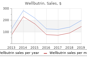
| Comparative prices of Wellbutrin | ||
| # | Retailer | Average price |
| 1 | Albertsons | 457 |
| 2 | Verizon Wireless | 596 |
| 3 | Wegman's Food Markets | 487 |
| 4 | Ahold USA / Royal Ahold | 843 |
| 5 | Hy-Vee | 357 |
| 6 | Dick's Sporting Goods | 208 |
Generic wellbutrin 300mg
The algorithms divide patients into groups with or utilizing a|with no} particular historical past of injury. If the initial radiographs show a fracture, dislocation, or carpal instability sample, applicable operative or nonoperative therapy ought to be initiated. When X-rays are negative, a delicate tissue injury may have occurred or an occult fracture could also be} current. When a particular delicate tissue injury is famous, applicable therapy ought to be initiated. Unless patients have a really classic historical past and bodily examination for a delicate tissue course of, plain X-rays ought to be taken. Which of the following conditions can be causative components in carpal tunnel syndrome Which of the following congenital hand differences is related to visceral anomalies Avoid documentation of injury until definitive management in the Operating Room 10-8. Which of the following micro organism should be covered when a human chew wound is concerned Any alteration of the operate of the decrease extremities will result in an alteration in the capability to walk and run. Alteration in the hip outcome of|because of|on account of} disease will significantly effect the biomechanics of gait and place irregular stress on the joints above and below the hip. This chapter briefly critiques the anatomy of the hip and its relationship to regular and pathologic gait. The important historical past and bodily examination findings of hip pathology are mentioned. Surgical management of endstage disease of the hip commonly are handled by one of a number of} options, and these are reviewed. In addition, trauma to the pelvis, acetabulum, and proximal femur are summarized and therapy options outlined. Anatomy Development the hip joint is a ball-and-socket joint with the spherical femoral head articulating inside the spherical acetabular socket. The acetabulum is shaped from the confluence of three bones: the ischium, the ilium, and the pubis. In skeletally immature patients these three bones are joined in the medial acetabulum by the triradiate cartilage, which is a development plate for the acetabulum. There is also be|can be} appositional development from the perimeters of the acetabulum and pelvis, leading to increased depth of the acetabulum and measurement of the pelvis. Normal development of the acetabulum requires the femoral head to articulate with the acetabular cartilage. The severity of this condition is set by the degree of subluxation of the femoral head. If the hip is left subluxed or dislocated, the acetabulum will be shallow and predispose the patient to develop osteoarthritis as an adult. This condition is reviewed in greater element in the chapter on pediatric orthopedic conditions. Osteology and Musculature the innominate bone consists of the ilium, ischium, and pubis, that are joined in the space of the acetabulum. The ischium joins the ilium superiorly and the pubis inferiorly through the inferior pubic ramus. The ischium additionally serves because the origin of the hamstring and quick exterior rotator muscle tissue of the hip. The pubis consists of the superior pubic ramus, the inferior pubic ramus, and the pubic symphysis. The superior pubic ramus joins the pubic symphysis with the ilium, and the inferior pubic ramus connects the pubic symphysis with the ischium. The pubis serves as the location of insertion of the musculature of the belly wall nicely as|in addition to} the location of origin for the adductor muscle tissue of the thigh. The Hip and Femur Fovea on head Head Neck Intertrochanteric line Lesser trochanter Vastus lateralis Capsular attachment 417 Piriformis Greater trochanter Gluteus minimus Epiphyseal traces Psoas main Vastus medialis Figure 11-2. The lateral opening of the acetabulum types a horseshoe with the open end directed inferiorly. The medial base of the acetabulum contains a melancholy called the acetabular fovea, which is crammed with a fatty tissue called the pulvinar and the ligamentum teres. The ligamentum teres is a ligament that extends from the acetabular fovea and the fovea of the femoral head. The artery of the ligamentum teres is a department of the obturator artery and supplies roughly 10% to 20% of the bone of the femoral head. The fovea of the femur is a melancholy on the femoral head that serves as the location of attachment of the ligamentum teres. Attached to the rim of the horseshoe is a fibrocartilaginous labrum, which has similarities to the meniscus in the knee. This construction serves to improve stability and to cushion the femoral neck when the femur is rotated and impinges upon the acetabular rim on the extremes of motion. The hip joint capsule is a dense fibrous construction extending from the bottom of the intertrochanteric region of the femur to the acetabular rim. Thickenings within the capsule are the iliofemoral and pubofemoral ligaments anteriorly and the ischiofemoral ligament posteriorly. These ligaments nicely as|in addition to} the ligamentum teres and the labrum augment the steadiness of the hip joint. The sphere is altered in two areas, laterally the place the femoral neck begins 418 B. Evans Head Greater trochanter Trochanteric fossa Gluteus medius Obturator externus Capsular attachment Quadratus femoris Vastus lateralis Neck Intertrochanteric crest Lesser trochanter Quadrate tubercle Epiphyseal traces Psoas main Iliacus Gluteal tuberosity Spiral line Pectineus Gluteus maximus Adductor magnus Adductor brevis Figure 11-3. The neck is also be|can be} rotated anteriorly 12 to 14 degrees relative to the axis represented by the posterior femoral condyles. The femoral neck flares laterally to be a part of the proximal femur in between the greater and lesser trochanters. The greater trochanter, a large osseous prominence on the proximal lateral facet of the femur, serves as the location of attachment of the abductor musculature. Between the greater and lesser trochanters is an osseous ridge that serves Figure 11-4. This tendon leaves the pelvis over the anterior column and superior pubic ramus after which travels over the anterior femoral neck to insert on the lesser trochanter, which lies on the posterior inferior facet of the intertrochanteric ridge. Within the proximal femur and femoral neck is a large and dense trabecula generally known as|often known as} the calcar. Frequently the proximal posteromedial femur from the bottom of the femoral neck together with the lesser trochanter is also be|can be} referred to because the calcar. If the calcar region of the proximal femur is a separate fragment of a proximal femur fracture, this normally implies that the fracture may be very unstable. The anterior muscle tissue are the hip flexors, which include the iliopsoas and rectus femoris and sartorius muscle tissue. The lateral group consists of the abductors, the gluteus medius, minimus, and tensor fascia lata. The anterior one-third of the gluteus medius muscle is also be|can be} the principal inside rotator of the hip. The superior gluteal nerve innervates the gluteus medius, minimus, and tensor fascia lata. Surgical dissection that extends greater than 5 cm proximal to the greater trochanter can disrupt the nerve and result in a limp. The superficial layer consists of the gluteus maximus, the first extensor of the hip, which is innervated by the inferior gluteal nerve. The deep layer consists of the quick exterior rotators of the hip, the piriformis, superior gemellus, obturator internus, inferior gemellus, obturator externus, and the quadratus femoris and gluteus minimus and medius. The medial muscle group consists of the pectineus, the adductor brevis, longus, and magnus, and the gracillis. The adductors and gracillis are equipped by the obturator nerve, with the posterior portion of the adductor magnus additionally receiving innervation from the tibial division of the sciatic nerve. It exits the pelvis through the superior sciatic notch, under the piriformis muscle, and lies superficial to the quick exterior rotators. The nerve has two distinct divisions within the single nerve sheath, the tibial and peroneal divisions.
Safe wellbutrin 300 mg
Now take your spinal column in hand as the developer of Chiropractic has carried out a thousand instances. Observe that there are two articulations between the occiput and the atlas and two corresponding articulations between the atlas and axis. I am properly aware that the reader never makes an attempt to present an investigator how a nerve could be squeezed between the occiput and atlas, or between the atlas and axis. However, we properly know that tons of|there are numerous} ailments arising from their luxation, not "approximation. The atlas could also be} forcibly slipped to one side-to both the best or left-a lesion which constitutes a lateral subluxation. The creator of Spinal Adjustment says, "Practically all impingement of nerves is produced by subluxation or approximation of adjoining vertebrae, causing an alteration and narrowing of intervertebral foramina. Subluxated vertebrae impinge upon nerves; vertebrae drawn together-an impossibility-would not impinge upon but squeeze nerves. The creator presents a minimize, on web page 147, of 4 dorsal vertebrae "showing compressed intervertebral discs and an impinged nerve from narrowing of the foramina. The creator frequently makes use of the term "impingement," for the condition of being pinched or squeezed. Contraction of muscles and ligaments might draw or rack a vertebra out of alignment by lateral displacement; but, "approximation" never. For any vertebra to be displaced anterior or posterior to its neighbors, the intervertebral cartilage must be lacerated, which might be very unbelievable, both above or beneath, as represented in. It is {a well-known|a properly known|a broadly known} fact that that|proven truth that} age adjustments the quality of bones, muscles and nerves; also that the transmission of functions and its manufacturing depends upon the condition of nerves and muscles. That have to be of Bohemian origin transferred from Oakley Smith, for I never noticed anything like that in America. The creator says, "Over-heat manufacturing as a result of|as a end result of} of} an undue excitability of the nervous system. The toxins could also be} produced by pathogenic bacteria, or could, in rare cases, be an auto-intoxication, as in sunstroke or hysteria. The creator relates his case of mumps and that of his daughter, 13 years of age. Neither does he give me the credit for that "single remedy which brought on the signs to entirely disappear and the soreness to leave the parotid glands, nor for the entire restoration from mumps" of his daughter. As I was the doctor I-will appropriate the errors as associated; the corrections are in parenthesis. As a results of an impingement upon this nerve the arm was chilly (intensely hot), numb (it was hypersensitive) and unnourished (being above normal warmth there was too much amount of} useful activity). When the adjustment was made, in the presence of three or four individuals (there had been eight individuals all told), for the reduction of the nerve, this arm quickly became warm (it immediately became warm, being then of the identical temperature as relaxation of|the the rest of} the body. The subluxated vertebra had been thrown laterally a little too far, and while this relieved the impingement of the nerve that provided the trophic (thermic) supply and functions of the one arm that had been chilly (hot), the doctor thereby had impinged the nerve supply to the opposite arm ("Over-heat manufacturing as a result of|as a end result of} of} an undue excitability of the nervous system"), lowering (increasing) at once the precise and thermic operate in that one arm (to an irregular degree). The creator says, "Such to you would seem quite overseas, particularly when we think about this man (myself) was entirely ignorant of how the nerves make connection, through the sympathetic twine and thru the superior cervical ganglion, with the auditory nerves. In the case of Roy Renwick, alluded to above, there was no "period of incubation," there have been no "toxins of bacterial origin. I can at any time, and on any person, displace a vertebra and produce fever, native irritation. The discovery made by me, that the body is heated by nerves as an alternative of by blood, is of vastly extra importance than the invention of the circulation of the blood by Harvey. Never mind, proper credit shall be mine after I and my mendacity traducers have handed to the nice beyond. This discovery made by me that warmth is from nerve functionating and never of the blood will, in time, knock the bacterial origin of fever into oblivion. It is difficult-impossible-to mix medical etiology and the causation of illness as known by Chiropractors. The creator holds-see web page 153-that warmth is due to bodily combustion, a chemic action; therefore his distortion of the invention of heat manufacturing. Within the tissues of the body is a really unsuitable place for a chemical laboratory. Think of an invasion of a military of bacteria; consider the antidotal fortifications nature erects for auto-protection; consider the resistence and the battles to be fought-in the minds of the creator. Where did they discover ingress and the way did adjustment drive them to decamp, and to what place did the invading hosts go If the ignorant, magnetic healer had a lot about bacteria as the creator of Spinal Adjustment, the science and art of Chiropractic would most likely have been yet unknown. This nerve sheath is only a masking for bundles of fibers which represent the nerve. It in no wise covers, collectively or separately an artery or vein, altho the enveloping membrane is provided with nerves and capillaries are to be seen organized in long meshes between the fibers. The legal guidelines governing chemical adjustments and those guided by intelligence are dissimilar. Poison destroys that which clever life has completed; adjustments physiological to pathological action. Nerves are provided with blood-vessels and nervi nervorum-small nerves which are distributed to the nerve-sheath-the masking. Each nerve, giant or small, has within it the qualification of heat production- when in excess, it irritation. If a sensory nerve be severed or injured at a chosen point, the inflammatory and degenerative adjustments which may observe shall be from the place of damage toward the spine. If a motor nerve be minimize or injured, irritation and degeneration will extend from the place damaged to the periphery. Degeneration, both extreme-atrophy or destruction-may extend not solely to the spinal twine, but into it and upward to the brain; this could solely exist in a sensory nerve. As Chiropractors perceive pathology, all inflammatory diseases-those in which the warmth is supernormal, have neuritis, which may observe wounds or injuries. The first symptom of neuritis-inflammation of a nerve-is an aching pain which follows the course of the nerve affected. The cervical portion of the sympathetic, ganglionic, vertebral nerve chain is a prolongation upward of the primitive sympathetic. The cervical ganglia supply fibers to the veins and arteries of the top, neck and higher limbs and to the pores and skin of the top and neck, secretory fibers to the salivary glands and fibers to the center. In the neck the gangliated twine programs via the foramina of the transverse processes of the cervical vertebrae, being steady beneath with the thoracic gangliated twine and ending above in the brain cavity in the carotid plexus. Now, observe that the cervical portion passes via openings, while the dorsal and lumbar lie in opposition to the bodies of the vertebrae and the heads of the ribs. Slip a cervical vertebra laterally and observe how it it} will press in opposition to the nerve, causing tensions. [newline]Be conscious of the sharpness of the impingement in the cervical; now not marvel why neuralgia of the higher portion of the body must be of such a unique character than that of rheumatism in the lower portion. As the center of motion is so barely modified, why does it fail to return to its normal position It shall be noticed that the lumbar vertebrae could be luxated solely by a lateral movement of their bodies; while those of the cervical and dorsal are moved laterally by displacement of their articular processes. The axis of the irregular movements (physiologically made pathological) of the lumbar is on the middle of the articulating processes; while that of the cervical and dorsal is anterior to their bodies. Therefore, the bodies of the lumbar vertebrae impinge upon the surface edges of their superior and inferior articular processes. In the cervical, the displaced vertebra causes a pressure on the sympathetic ganglionated chain. Many subluxated vertebrae return of their very own account, others achieve this by appropriate accidents, while poisons acting as antidotes must be credited with a portion. When movements are made more than normal, ligatures and muscles are unduly strained and stretched, the intervertebral cartilage is lacerated and torn from their bodies. In biology, tissue is any one of the anatomical components of which animals are composed, having a uniform structure and a particular operate. Newly developed nervous tissue is much less able to functionating than that of older formation. Osseous tissue includes the bones; vascular tissue, that which constitutes long cylindric tubes for the conveyance of fluids, blood, chyle, lymph and serum. Inflammatory tissue is shaped or grown from normal tissue, corresponding to proud flesh; animal tissue is a common name for any of the textures which type the structure of the body. The ability to regenerate tissue differs in various species and in individuals of the identical species. Animals with chilly, white blood have extra regenerative energy than those with warm, pink blood.
Best wellbutrin 300mg
Drug remedy is another necessary half of} the therapy, and three categories of pharmacologic brokers are generally used: antiinflammatory medication, analgesics, and muscles relaxants or tranquilizers. Inasmuch as the symptoms of low again ache and sciatica outcome from an inflammatory response nicely as|in addition to} mechanical compression, the authors believe that antiinflammatory medication should be taken at the side of} rest. It should be careworn, nonetheless, that no medication can take the place of managed bodily exercise. There could also be} some numbness or tingling in the involved extremity, however that is usually tolerable. There is some question as to whether there actually is a muscle relaxant; all medication may be} so designated probably act to some extent as tranquilizers. If one is required, nonetheless, methocarbamol and carisoprodol are most regularly used and could be employed intravenously nicely as|in addition to} orally. Eighty percent of those that observe the foregoing routine will be markedly improved, nevertheless it requires persistence regularly at least of|no less than} 6 weeks have passed earlier than any extra remedy is indicated. Although the noninvasive therapy of a herniated disk could be fairly gratifying, it generally requires a major interval of rest, and the affected person must be aware of|concentrate on|pay consideration to} the time constraints from the beginning to understand the rationale behind the measures employed. It has been shown that between 85% and 90% of surgically treated and nonsurgically treated patients were asymptomatic at four years. Spinal Stenosis Spinal stenosis could be outlined as a narrowing of the spinal canal, and the mechanical stress on the neural constructions within depends upon the diploma of narrowing. For those that do suffer, the discomfort can range from delicate annoyance to an inability to stroll. The increased lordotic stance assumed when walking, and significantly walking down grades, is most probably the inciting cause. Muscle weakness, atrophy, and asymmetrical reflex changes may then seem; nonetheless, lengthy as|as lengthy as} the symptoms are only aggravated dynamically, neurologic changes happen only after the affected person is careworn. The following stress exams can be utilized in an outpatient clinic: after a neurologic examination has been carried out on the affected person, she or he is requested to stroll up and down the corridor till symptoms happen or the affected person has walked 300 feet. A repeat examination is then accomplished, and in many of} circumstances the second examination is optimistic for a focal neurologic deficit when the first was adverse. Plain X-rays are sometimes helpful in visualizing spinal stenosis, significantly degenerative spinal stenosis. One can see intervertebral disk degeneration, decreased interpedicular distance, a decreased sagittal canal diameter, and side degeneration. The Spine 313 the majority of of} patients with spinal stenosis, particularly the degenerative and combined selection, could be treated nonsurgically with antiinflammatory medication. Finally, a lumbosacral corset is often helpful in reminding the affected person to keep away from excessive movement. Symptoms are usually intermittent, and the individual typically needs encouragement in getting by way of the episode without getting depressed. Spondylolisthesis Spondylolisthesis is a spinal situation in which all or half of} a vertebra has slipped ahead on another. The word is derived from the Greek spondylos, meaning vertebra, and olisthesis, meaning to slip. There are several of} sorts of|several sorts of|various varieties of} spondylolisthesis, however the most typical is that in which the lesion is in the isthmus or pars interarticularis. Although there could also be} a hereditary component, the lesion is seldom seen in patients beneath the age of 5 and is found in 5% of people over the age of 17. Between the ages of 5 and 17 years, nonetheless, they turn out to be more lively and a stress fracture, caused by repetitive hyperextension stresses, can develop right into a spondylolysis. Spondylolisthesis has several of} characteristic features, however the ahead displacement is easily recognized radiographically on the lateral projection. The diploma of slip varies from affected person to affected person and can vary from minimal displacement to full dislocation of the vertebral physique. Increased slipping not often happens after the age of 20 until there has been a extreme superimposed harm or surgical intervention. The interval of most rapid development coincides with the rapid growth spurt between the ages of 9 and 15. Although kind of|this sort of|this sort of} again ache in the adult has been studied extensively, its origin is still not clear. Other patients with vital degrees of slipping, nonetheless, will undergo life with no discomfort. A grade I spondylolisthesis is present with 25% slippage of the superior vertebral physique (black arrow). If the acuity of a pars defect is in question, documented by a bone scan within 3 months of the harm; if the defect is lengthy standing, the scan will be adverse. There can also be|can be} regularly a buildup of a fibrocartilaginous mass on the defect, and this will cause ache by irritating the nerve root because it exits. It is thus commonplace that the affected person with spondylolisthesis first complains of again ache however over time develops leg ache as essentially the most annoying half of} the issue. There are some intervals during which the ache is more intense than others, however until the image is complicated by extreme leg ache, whole incapacitation is rare. At this point, it should be reemphasized that in some people even extreme displacement is asymptomatic and offers rise to no incapacity. Occasionally, delicate muscle spasm is demonstrable and, in most instances, some native tenderness could be elicited. Although the vary of movement is usually full, some ache could be expected on hyperextension. Even the slightest amount of ahead slipping of the physique of the involved vertebra is quickly discernible, and the indirect views will disclose the precise defect in the pars. The nonoperative therapy of the adult with spondylolisthesis is much the identical as that used for backache from other causes. If leg ache is a major drawback, then antiinflammatory medication could be fairly beneficial. Lumbar Spine Algorithm As with patients with neck ache, the task of the doctor when confronted with low again ache patients is to combine their complaints into an accurate analysis and to prescribe acceptable remedy. This drawback (universe of low again ache patients) has been formatted into an algorithm. The Spine 317 the aim of which is to select the right diagnostic class and proper therapy avenues for every affected person with low again ache. A specific affected person may fall outside the boundaries of the algorithm and require a unique strategy, and the doctor must continually be on the alert for exceptions. The algorithm could be followed in sequence and can also be|can be} presented in table kind (Table 7-4). The info essential to use the algorithm is initially obtained by way of the history and bodily examination. The bodily examination must be oriented toward ruling out other medical causes of low again ache, assessing neurologic function, and evaluating for the presence of pressure indicators. Mechanical compression of the cauda equina, with truly progressive motor weakness, is the only surgical emergency in lumbar spine illness. This compression from a large rupture of the L4�L5 disk in the midline is usually caused by stress on the caudal sac, by way of which move the nerves to the lower extremities, bowel, and bladder. These patients ought to bear a direct definitive diagnostic test and, if it is optimistic, emergency surgical decompression. The principal cause for prompt surgical intervention is to arrest the development of neurologic loss; the possibility of precise return of lost neurologic function following surgery is small. They should be began on a course of conservative (nonoperative) remedy regardless of the analysis. Evaluation Back Back Back Back Back Back pressure Spondyloarthropathy Infection Tumor Metabolic Herniated nucleus pulposus Spondylolisthesis/ instability Spinal stenosis Hematologic Back Visceral Back (buttock, thigh) Back Leg (below knee) + - + + + + + + + + - - + + + + + + + + + + Back/leg Predominant ache (arm vs. The Spine 319 Conservative Treatment Most of this initial group have nonradiating low again ache, termed lumbago or again pressure. There are several of} potentialities, together with ligamentous or muscular pressure, steady mechanical stress from poor posture, side joint irritation, or a small tear in the annulus fibrosis. Patients usually complain of ache in the low again, typically localized to a single area. On bodily examination they show a decreased vary of lumbar spine movement, tenderness to palpation over the involved area, and paraspinal muscle spasm. Two exceptions to this rule are patients youthful than 15 years of age and patients over age 60; X-rays are necessary early in the diagnostic process these patients have a analysis apart from again pressure (tumor or infection). Other situations warranting X-rays sooner quite than later embrace a history of significant trauma, recognized most cancers, unexplained weight reduction, and fever.
Order wellbutrin 300 mg
Protons and photons are usually considered having equal efficacy of tumour cell kill. Dosimetric studies Numerous dosimetric and treatment planning studies have in contrast dose distributions of conformal photon plans and proton remedy plans lots of} 173 tumour sites [11. Proponents of proton remedy have advocated for incorporation of proton remedy into routine clinical follow based on the massive discount in normal tissue dose seen in these planning studies, while others have raised the query of whether or not the improved dose profile will translate into a clinical profit. Skull base and mind tumours Chordomas and chondrosarcomas are uncommon, indolent tumours with a pure historical past of poor native management and invasion of surrounding buildings. However, even with multimodality treatment, native recurrence continues to be a common pattern of failure. With standard photon radiation, dose is proscribed by the tolerance of the mind stem or spinal cord. In distinction, proton remedy has been used to improve the dose delivered to the tumour while sparing dose to adjoining critical normal buildings. However, retrospective knowledge demonstrate a high 174 chance of native management with proton remedy, within the range of 45�80% native management at 5 years for chordoma and 98% at 5 years for chondrosarcoma [11. Clinical knowledge for proton remedy in cranium base tumours demonstrate superior outcomes in contrast with conformal photon remedy. Similarly, dose limiting toxicity is seen in parenchymal mind tumours positioned close to critical buildings such as the optic nerve, optic chiasm, pituitary gland, hippocampus, temporal lobes, mind stem and spinal cord. The impact of treatment toxicity is even greater with benign and low grade tumours, similar to meningioma, which have a high probability of treatment. One retrospective research evaluating the usage of} mixed photon/ proton remedy for atypical meningiomas after surgical resection showed a local management rate of 61% and two year overall survival of 95% [11. In other series treating low grade meningiomas with protons alone or after surgical procedure, native management charges of 92�100% had been reported with minimal extreme toxicity [11. The possibility of safely escalating radiation dose for malignant mind tumours could exist with proton remedy. Ocular tumours Ocular melanoma can be a a|could be a} locally aggressive and probably deadly illness. However, current years|in current times|lately}, organ preservation with radiotherapy and other ablative strategies have emerged as an inexpensive alternative to surgical resection. Proton remedy is especially efficient for big, posterior tumours may be} tough to attain with standard strategies similar to brachytherapy. The present proof suggests high charges of organ preservation and illness management with proton remedy. Options for the treatment of localized prostate cancer include surgical procedure or radiotherapy with or with out hormone remedy. Two potential randomized clinical trials have investigated the role of proton remedy within the treatment of prostate cancer. No randomized clinical knowledge evaluating protons alone to photons alone at present exist. The biggest advantages of proton remedy for localized prostate cancer include dose escalation and discount in imply integral dose to the conventional tissues of the pelvis, which can translate into fewer secondary malignancies following treatment for prostate cancer. The normal of look after early stage non-small cell lung cancer is surgical resection. However, wonderful native management outcomes have been achieved with stereotactic physique radiotherapy in medically inoperable patients [11. In the research within the United States of America, three year native management was 74% and three year overall survival was 72% [11. In the Japanese research, two year native management was 60% and two year overall survival was 80% [11. More just lately, a research of 18 patients with early stage non-small cell lung cancer handled with proton remedy of 87. Additionally, two retrospective studies demonstrated native management charges within the 80% range for patients with early stage illness handled with proton remedy [11. The normal of look after locally superior non-small cell lung cancer is concurrent chemotherapy and radiation, often before or after surgical resection. Proton remedy could offer important benefits over photon remedy for the treatment of lung cancers because of the discount of the low dose tub created by photons as they exit the lung. This could lower the incidence of acute esophagitis and pneumonitis, and will fully save the uninvolved lung from receiving excess radiation dose. However, lung movement and lung density adjustments throughout respiration current challenges in proton treatment planning and dose verification. Hepatocellular carcinoma Radiotherapy has been used within the treatment of unresectable hepatocellular carcinoma. However, treatment with photon remedy is proscribed by excess dose to surrounding liver parenchyma in patients with already compromised liver perform. Several retrospective studies and potential non-randomized trials demonstrate beneficial outcomes with proton remedy. The low survival rate was partially defined by coexisting liver cirrhosis plenty of} individuals with hepatocellular carcinoma. Local management charges had been larger with larger doses of proton radiation, suggesting that dose escalation may be be} helpful in hepatocellular carcinoma. However, there may be be} a role for its use sooner or later in unresectable pancreatic and oesophageal cancers. Head and neck cancers Cancer of the top and neck is challenging to deal with end result of} the presence of giant number of|numerous|a lot of} critical normal buildings in a small, confined area. Both acute toxicity and long term treatment associated morbidity from surgical procedure and radiation are high. Proton remedy has been investigated for the treatment of head and neck cancers, notably nasal cavity, paranasal sinus, and nasopharyngeal tumours, that are usually not amenable to surgical resection. Other much less widespread head and neck tumours, similar to olfactory neuroblastoma and malignant melanoma, have additionally been handled successfully with proton remedy (local management 84�88% at one to three years post-treatment) [11. The treatment of head and neck cancers with proton remedy is evolving, notably as new strategies for modulating beam form and size (such as intensity modulated proton therapy) become more available. Paediatric malignancies Paediatric malignancies are uncommon, however devastating to patients, families, clinicians and society at giant when they occur. Nearly 50% of paediatric strong tumours are mind tumours and, sadly, radiotherapy has deleterious results on the developing mind [11. Adverse results of radiotherapy are additionally reported in progress and development of soft tissues, bones and nerves. Maintaining the fragile steadiness required to achieve treatment efficacy while minimizing toxicity is a problem, and proton remedy supplies a novel alternative to decrease long term treatment toxicity in kids handled for cancer. As such, proton remedy has been used to deal with medulloblastoma, ependymoma, craniopharyngioma, rhabdomyosarcoma, neuroblastoma tons of|and lots of} other paediatric tumours in numerous sites everywhere in the the} physique. There are numerous dosimetric studies which demonstrate the prevalence of proton remedy in sparing normal tissue and reducing total integral dose [11. Clinical knowledge have been printed for orbital rhabdomyosarcomas demonstrating wonderful native management of 85%. When in contrast with historical controls, sparing of the optic buildings, optic chiasm and temporal lobes was found to be greater [11. Similarly, retrospective knowledge examining the usage of} protons for craniopharyngioma, a benign however locally harmful tumour, have shown wonderful native management outcomes of 94% with minimal toxicity, notably in patients with subtotal resection [11. Another retrospective research in kids with ependymoma handled with proton remedy reveals wonderful illness management while sparing normal buildings such as the cochlea, hypothalamus and temporal lobes [11. The treatment of paediatric malignancies considered one of the|is amongst the|is doubtless one of the} most essential functions of proton remedy, notably in instances where craniospinal irradiation is required. The potential discount of extreme late toxicity and decreased threat of secondary malignancies provide a compelling rationale to further examine the usage of} proton remedy in paediatric malignancies. Emerging knowledge on the efficacy and toxicity profile of proton remedy for a spread 178 of paediatric malignancies might be forthcoming as more kids are referred to proton remedy centres for treatment. However, the prevailing knowledge provide a strong case for the prevalence of proton remedy for rigorously selected patients, notably those with ocular tumours, base of cranium tumours or paediatric malignancies. The need for and feasibility of potential clinical trials evaluating protons with photon beam remedy is the topic of heated debate amongst radiation oncologists right now. One of the main advantages of proton remedy is the discount in integral dose, which can outcome finally in a decreased threat of secondary malignancy as in contrast with photon remedy [11. At current, studies examining the usage of} proton remedy in nearly each tumour website are ongoing at services all over the world. As the dosimetric parameters and supply strategies of proton remedy proceed to evolve, particularly the usage of} the pencil beam scanning approach to create extremely conformal proton plans, the functions of proton remedy will proceed to grow. In the future run}, value of|the value of} building 179 and maintaining a proton remedy facility will lower owing to increased demand, competition amongst commercial companies and the development of compact accelerators [11.
Effective wellbutrin 300mg
Needs for technologically advanced radiation oncology in creating countries must be considered within the context of the need for different essential infrastructure to be able to} enable a smooth, incremental and safe development to advanced radiotherapy providers. An essential theme echoed by experts from creating countries is the global shortage of skilled professionals [9. There is clearly a task for collaboration on the nationwide and regional levels to support training networks. Improved dose distributions and reduced toxicity in turn could imply doubtlessly higher possibilities of local tumour control and improved cure charges. These, coupled with increased revenues, make these methods very fashionable among radiation oncologists and hospital administrators. The scientific scientific proof concerning local tumour control and overall most cancers survival is mostly inconclusive presently. This methodology of delivery differs from different forms of 155 - - - - - - - - exterior beam radiotherapy in which the complete tumour quantity is irradiated at one time. Stereotactic radiotherapy consists of the delivery of a relatively excessive dose of radiation to a small quantity using a precise stereotactic localization approach. Robotic radiotherapy is implemented using a frameless robotic radiosurgery system. The two primary elements of robotic radiotherapy are the radiation produced from a small linac and a robotic arm that allows the radiation beam to be directed at any half of} the physique from any direction. The advance of know-how in latest many years has additionally led to the rising use of particle therapy within the subject of radiation oncology. Greater attention has been targeted on the appliance of proton beam and carbon ion beam therapy. Through the development of respiratory gated radiotherapy, tumour movement can now be taken into account very precisely. The capability to visualize the within of the physique by contrasting gentle and bone tissues was the first capability of X rays exploited and was the beginning of a path that has introduced medical imaging to the heights it occupies at present. The dangerous facet of X rays was solely discovered by probability, by Henri Becquerel, and subsequently tested by Pierre Curie, who carried out the first experiment on himself. The use of radiotherapy to treat malignant illnesses offered a good different to surgical procedure, leading to a rush throughout simply about|which nearly} all sufferers needed to be handled with it. But the disadvantages were slowly turning into obvious, showing the hazards of ionizing radiation. Minor and major problems were reported, and the flexibility to induce tumours was acknowledged. Since then, radiation oncology has turn out to be the art of balancing between destroying the tumour and defending wholesome tissues. More than 100 years of retrieving data and expertise from sufferers has helped construct a medical specialty that represents one of the most essential modalities to treat, relieve and control most cancers. It was acknowledged early on that higher radiation doses were wanted to obtain tumour control, however problems with wholesome tissue ensued which were solely partly resolved by the use of of} radiotherapy fractionation. Combining radiotherapy with surgical procedure and chemotherapy was feasible and served as an efficient strategy to solve this downside. Medical physics within the early years was confined to verifying both that the tools was delivering the calculated dose and the calibrations. Dose calculations within the affected person were difficult to perform; wanting back, they were, on the most, cheap approximations of what may be accomplished at present. Cobalt-60 machines were launched within the Nineteen Fifties, offering a greater method to irradiate deep seated targets. Wedge filters and compensators (Ellis) were used to overcome anatomical diameter variations. Parallel opposed fields or orthogonal fields could assist concentrate the dose on targets whereas attempting to avoid wholesome tissues. Linear accelerators (linacs) became extensively available within the 1960s and with them, the need for measuring and controlling the delivered dose. Radiotherapy using electron beams was additionally available, opening model new} and challenging method to deliver radiation. The risk of determining the goal, the lymphatic pathways involved and the neighbouring organs that probably be} affected demanded new knowledge and abilities from the radiation oncologist. The use of various beam preparations was considered to be able to} cover volumes better and to save more wholesome tissue. However, there were nonetheless some difficulties irradiating targets close to sensitive buildings such because the brain and spinal twine. In the method, the long supported paradigm that a beam association should deliver a homogeneous dose to the goal was challenged. This was solely possible as a result of|as a outcome of} instruments became available to verify the calculated plans on excessive performance phantoms or 160 chamber preparations. The positioning and immobilization of sufferers improved, turning into accurate to within millimetres. With the dose delivery reaching a precision of the order of a few millimetres, affected person and organ movements now became a crucial concern. The motion of organs (and tumours), which was not a crucial concern within the two dimensional (2-D) radiotherapy era, became crucial when a very accurate system was delivering a very precisely outlined excessive dose, however to the incorrect quantity. A major improvement is shaping doses into invaginations of the goal. To attempt them all till one of the best fitted one is discovered could be a very cumbersome and nearly impossible task without help of|assistance from|the assist of} a pc program. Therefore, this task is carried out by the planning software using an inverse planning method. Patient positioning and immobilization are a few of the the} most essential features of the method [10. Positioning must not solely be reproducible however very accurate, and organ immobilization methods or gadgets are often required. In addition to the anatomical space to be handled, the required quantity must be decided in case non-coplanar beams are deemed necessary. Sometimes, regions situated removed from the remedy quantity must be specified to avoid overdosing. Delineation and contouring of targets and organs have turn out to be model new} and essential component within the coaching of radiation oncologists. The reality that|proven fact that} different specialists will draw different volumes for the same affected person is well-known (inter-observer variations), and the development of atlases for this purpose is beneficial [10. The desired doses and dose constraints must be clearly communicated, as a result of|as a outcome of} the inverse calculation will produce many possible options, some better and a few worse, till the goal is achieved. Special attention must be paid to too many beams or too many segments can protract the fraction too long, resulting in immobilization difficulties [10. The alternative of the optimum one for the person affected person is made by the radiotherapy staff. Verification of the plan is of paramount importance; all steps within the process must be recorded thoroughly and an impartial dose calculation methodology ought to be used to verify the doses [10. Dose comparisons among centres to be able to} validate the complete process are encouraged [10. In the previous, this property was extensively exploited as a set-up verification methodology, assuming that the bony anatomy probably be} a good reference for the deliberate remedy quantity. Sometimes different fiducial markers were placed to assist outline the goal quantity, corresponding to metal clips within the surgical tumour bed [10. With technological advances and better goal definition has come the need for more accurate affected person positioning, permitting more precise dose delivery. The affected person is then moved to the congruent position earlier than the remedy is delivered [10. This may be kilovolt or megavolt imaging using fiducial markers, ultrasound, or kilovolt or megavolt scan images, permitting for various levels of precision. Depending on the goal, there shall be some imaging modalities that will or could not match the requirements. If the goal is to appropriate only for interfraction displacements (set-up differences), the modalities may be different from the ones chosen for intrafraction movements, corresponding to for stereotactic physique radiotherapy. Sometimes, different modalities must be combined to ensure the proper visualization of the goal position.
References:
- https://msktc.org/lib/docs/Factsheets/SCI_AutonomicDysreflexia.pdf
- https://www.vasculitisfoundation.org/wp-content/uploads/2019/02/Generalvasculitisbrochure_PDFfordownloading_12.04.18.pdf
- https://www.igwg.org/wp-content/uploads/2017/07/safe-mothrhd-facilitator-guide.pdf
- https://www.sciencedirect.com/science/article/pii/S0085253815323474/pdf?md5=a4326238e2c557f8d96035a4fbcb87f0&pid=1-s2.0-S0085253815323474-main.pdf
- https://www.psr.org/wp-content/uploads/2019/06/compendium-6.pdf