
.png)
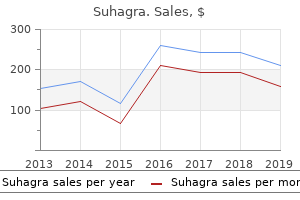
Purchase 100mg suhagra
Without their strong engagement, the goal of free entry to high-quality textbooks would stay only a dream. The William and Flora Hewlett Foundation has been making grants since 1967 to assist remedy social and environmental issues at home and around the world. The Foundation concentrates its assets on actions in schooling, the environment, international growth and inhabitants, performing arts, and philanthropy, and makes grants to help deprived communities within the San Francisco Bay Area. Kazanjian Economics Foundation Inc, in 1949 as a philanthropic, nonpolitical academic organization to help efforts that enhanced economic understanding. Guided by the assumption that every life has equal worth, the Bill & Melinda Gates Foundation works to assist all folks lead wholesome, productive lives. In the United States, it seeks to significantly improve schooling so that all one|that every one} young folks have the chance to attain their full potential. The Maxfield Foundation helps tasks with potential for prime impact in science, schooling, sustainability, and different areas of social importance. We help the creation, sharing, and proliferation of simpler, extra affordable academic content material by leveraging disruptive technologies, open academic assets, and new fashions for collaboration between for-profit, nonprofit, and public entities. The Bill and Stephanie Sick Fund helps innovative tasks within the areas of Education, Art, Science and Engineering. Animal Structure and Function Chapter 33: the Animal Body: Basic Form and Function. This textbook has been created with a number of} objectives in mind: accessibility, customization, and pupil engagement-all while encouraging science college students toward high levels of academic scholarship. Instructors and college students alike will discover that this textbook presents a strong foundation in biology in an accessible format. About OpenStax OpenStax is a non-profit organization committed to improving pupil entry to quality studying supplies. Unlike traditional textbooks, OpenStax assets stay on-line and are owned by the neighborhood of educators utilizing them. Through our partnerships with corporations and foundations committed to lowering prices school kids|for faculty students}, OpenStax is working to improve entry to greater schooling for all. OpenStax is an initiative of Rice University and is made attainable by way of the generous help of a number of} philanthropic foundations. Customization OpenStax studying assets are designed to be custom-made for each course. Our textbooks present a strong foundation on which instructors can build, and our assets are conceived and written with flexibility in mind. Instructors can choose the sections most relevant to their curricula and create a textbook that speaks directly to the needs of their courses and pupil physique. Teachers are encouraged to broaden on present examples by adding unique context by way of geographically localized functions and topical connections. Simply choose the content material most relevant to your present semester and create a textbook that speaks directly to the needs of your class. Biology is organized as a group of sections might be} rearranged, modified, and enhanced by way of localized examples or to incorporate a selected theme of your course. This customization characteristic will assist convey biology to life for your college students and will ensure that that|be sure that} your textbook truly reflects the objectives of your course. Cost Our textbooks can be found at no cost on-line, and in low-cost print and e-book editions. About Biology Biology is designed for multi-semester biology courses for science majors. It is grounded on an evolutionary basis and contains exciting features that spotlight careers within the biological sciences and a daily basis} functions of the ideas at hand. Instructors can customise the e-book, adapting it to the approach that works finest in their classroom. Biology additionally contains an innovative artwork program that comes with crucial pondering and clicker questions to assist college students understand-and apply-key ideas. In developing Biology, we listened to tons of of General Biology instructors who readily offered suggestions about their courses, college students, challenges, and hopes for innovation. But extra importantly, these lecturers instructed enhancements for the textbook, which might ultimately result in extra meaningful and memorable studying experiences school kids|for faculty students}. We additionally try to make biology, as a discipline, interesting and accessible to college students. The pedagogical choices, chapter preparations, and studying objective achievement had been developed and vetted with the suggestions of one other 100 reviewers, who completely read the material and provided detailed crucial commentary. Our opening unit introduces college students to the sciences, together with the scientific methodology and the basic ideas of chemistry and physics that present a framework within which learners comprehend biological processes. Students will acquire strong understanding of the constructions, capabilities, and processes of the most basic unit of life: the cell. The core ideas of evolution are mentioned on this unit with examples illustrating evolutionary processes. Additionally, the evolutionary basis of biology reappears all through the textbook in general dialogue and is strengthened by way of special call-out features highlighting specific evolution-based topics. The variety of life is explored with detailed study of various organisms and dialogue of emerging phylogenetic relationships. This unit strikes from viruses to living organisms like bacteria, discusses the organisms previously grouped as protists, and devotes multiple of} chapters to plant and animal life. Our plant unit completely covers the basic information of plant life important to an introductory biology course. An introduction to the shape and function of the animal physique is adopted by chapters on specific physique techniques and processes. This unit touches on the biology of all organisms while maintaining an attractive focus on human anatomy and physiology that helps college students hook up with the topics. Ecological ideas are broadly lined on this unit, with features highlighting localized, real-world issues of conservation and biodiversity. Pedagogical Foundation and Features Biology is grounded on a strong scientific base and designed to assist college students perceive the ideas at hand. Throughout the text, one can explore features that have interaction the scholars in scientific inquiry by taking chosen topics a step additional. Our features embrace: Evolution Connection features uphold the importance of evolution to all biological study by way of discussions like "The Evolution of Metabolic Pathways" and "Algae and Evolutionary Paths to Photosynthesis. Features embrace "Determining the Time Spent in Cell Cycle Stages" and "Testing the Hypothesis of Independent Assortment. Everyday Connection features tie biological ideas to emerging points and discuss science phrases of|when it comes to|by way of} a daily basis} life. Topics embrace "Chesapeake Bay" and "Can Snail Venom Be Used as a Pharmacological Pain Killer Biology additionally incorporates hyperlinks to relevant animations and interactive workouts that assist convey biology to life school kids|for faculty students}. Link to Learning features direct college students to on-line interactive workouts and animations to add a fuller context and examples to core content material. Senior Contributing Authors Yael Avissar Jung Choi Jean DeSaix Robert Wise Connie Rye Rhode Island College Georgia Institute of Technology Cell Biology Genetics Animal Physiology Plant Biology General Content Lead University of North Carolina at Chapel Hill Evolution University of Wisconsin, Oshkosh east Mississippi Community College Vladimir Jurukovski Suffolk County Community College Contributing Authors and Reviewers Julie Adams Summer Allen James Bader David Bailey Mark Belk Nancy Boury Lisa Bonneau Mark Browning Sue Chaplin George Cline Deb Cook Diane Day Frank Dirrigl Waneene Dorsey Nick Downey Rick Duhrkopf Kristy Duran Stan Eisen Brent Ewers Aurora University Brown University Case Western Reserve University St. Norbert College Brigham Young University Iowa State University Metropolitan Community College - Blue River Purdue University University of St. Adaptive tools present college students with a private, adaptive studying expertise permit them to} build their proficiency on topics and use their study time most successfully. Biology Powerpoint Slides (faculty only) the PowerPoint slides openstaxcollege. SimBio is finest recognized for his or her EcoBeaker and EvoBeaker suites of simulated ecology and evolution laboratories that guide college students by way of the "discovery" of important ideas by way of a mix of|a mixture of} structured and open-ended experimentation on simulated techniques. In response to well-liked demand, SimBio has begun applying the same powerful approaches to topics in cell biology, genetics, and neurobiology. The first types of life on Earth are thought to have been microorganisms that existed for billions of years within the ocean earlier than vegetation and animals appeared. The mammals, birds, and flowers so acquainted to us are all comparatively latest, originating one hundred thirty to 200 million years ago.
Trusted 100mg suhagra
X-rays confirmed that bone destruction had already unfold to the tarsal bones, and after 2 years of futile therapy the foot had to be amputated. Diagnosis is usually delayed and infrequently involves specialised microbiological investigations to determine the organism. The definitive host is the dog or some other carnivore that carries the tapeworm in its bowel. Segments of worm and ova pass out in the faeces and are later ingested by one of many intermediate hosts � usually sheep or cattle or man. Here the larvae are carried through the portal circulation to the liver, and infrequently past to different organs, where they produce cysts containing numerous scolices. Infested meat is then eaten by canine (or humans), giving rise to a new new} technology of tapeworm. Scolices carried in the blood stream sometimes settle in bone and produce hydatid cysts that slowly enlarge with little respect for cortical or epiphyseal boundaries. The bones mostly affected are the vertebrae, pelvis, femur, scapula and ribs. Infestation generally starts in childhood however the cysts take so lengthy to enlarge that scientific signs and signs might not turn into apparent quantity of} years}. Imaging X-rays present solitary or multiloculated bone cysts, however solely moderate enlargement of the cortices. In the spine, hydatid illness might contain adjoining vertebrae, with large cysts extending into the paravertebral delicate tissues. Diagnosis Hydatid illness must be included in the differential prognosis of benign and malignant bone cysts and cystlike tumours. The anthelminthic drug albendazole is moderately efficient in destroying the parasite. The indications for surgical procedure are persevering with enlargement or unfold of the lesion, a danger of fracture, invasion of soppy tissues and strain on necessary buildings. Radical resection, with the margin at least of|no less than} 2 cm past the cyst, is extra certain, but in addition far more difficult. In a long bone the space can generally be full of a tumour-prosthesis, to include an arthroplasty if needed. Large cysts of the vertebral column, or the pelvis and hip joint, are significantly tough to handle in this method and in some instances surgical excision is solely impractical or unimaginable. The changing epidemiology of acute and subacute haematogenous osteomyelitis in children. A histological research of acute haematogenous osteomyelitis following physeal injuries in rabbits. Individuals with these ailments probably to|are inclined to} die youthful than their friends end result of|because of|on account of} the consequences of chronic inflammation. Many � perhaps all � are because of of} a defective immune reaction resulting from a mixture of environmental exposures towards a background of genetic predisposition. Rheumatoid arthritis is a systemic illness and changes could be widespread in quantity of|numerous|a selection of} tissues of the physique. Chief among these is early ischaemic coronary heart illness secondary to the consequences of inflammation on the cardiovascular system. Both the prevalence and the scientific expression differ between populations; the illness is extra frequent (and usually extra severe) in Caucasians dwelling in the urban communities of Europe and North America than in the rural populations of Africa. This might suggest that a particular antigen that matches into taking part in} a component. However, a great deal is now identified concerning the action is initiated, numerous local factors come into play and result in a progressive enhancement of the immune response. Immune complexes are deposited in the synovium and on the articular cartilage, where they appear to increase the inflammatory process. This combination of things results in depletion of the cartilage matrix and, eventually, harm to cartilage and underlying bone. Vascular proliferation and osteoclastic activity, most marked at the edges of the articular floor, might contribute additional to cartilage destruction and peri-articular bone erosion. There is thickening of the capsular buildings, villous formation of the synovium and a cell-rich effusion into the joints and tendon sheaths. Although painful, swollen and tender, these buildings are nonetheless intact and cellular, and the disorder is potentially reversible. Stage 3 � destruction Persistent inflammation causes Pathology Rheumatoid arthritis is a systemic illness however probably the most characteristic lesions are seen in the synovium or inside rheumatoid nodules. The synovium is engorged with new blood vessels and packed full of inflammatory cells. In anyone joint features of various phases could be occurring simultaneously and even when joints are very badly destroyed the ongoing inflammation can con- joint and tendon destruction. Articular cartilage is eroded, partly by proteolytic enzymes, partly by vascular tissue in the folds of the synovial reflections, and partly because of of} direct invasion of the cartilage by a pannus of granulation tissue creeping over the articular floor. At the margins of the joint, bone is eroded by granulation tissue invasion and osteoclastic resorption. Similar changes occur in tendon sheaths, inflicting tenosynovitis, invasion of the collagen bundles and, eventually, partial or complete rupture of tendons. A synovial effusion, typically containing copious amounts of fibrinoid materials, produces swelling of the joints, tendons and bursae. Stage four � deformity the mixture of articular destruction, capsular stretching and tendon rupture leads to progressive instability and deformity of the joints. The inflammatory process usually continues however the mechanical and functional results of joint and tendon disruption now turn into vital. Nodules occur beneath the skin (especially over bony prominences), in the synovium, on tendons, in the sclera and in most of the viscera. Lymphadenopathy Not solely the nodes draining infected joints, but in addition these at a distance such as the mediastinal nodes, could be affected. This, nicely as|in addition to} a gentle splenomegaly, is due to of|as a result of} of} hyperactivity of the reticuloendothelial system. Involvement of the skin, together with nailfold infarcts, is frequent however organ infarction can occur. It additionally be} because of of} a generalized myopathy or neuropathy, however exclude spinal wire illness or wire compression because of of} vertebral displacement (atlantoaxial subluxation). Sensory changes additionally be} half of} a neuropathy, however localized sensory and motor signs also can result from nerve compression by thickened synovium. Visceral illness the lungs, coronary heart, kidneys, gastrointestinal tract and mind are generally affected. In the early phases the picture is mainly that of a polysynovitis, with soft-tissue swelling and stiffness. Typically, a girl of 30�40 years complains of ache, swelling and lack of mobility in the proximal joints of the fingers. Another basic feature is generalized stiffness after periods of inactivity, and particularly after rising from bed in the early morning. If the bigger joints are involved, local warmth, synovial hypertrophy and intra-articular effusion additionally be} extra obvious. Movements are often limited however the joints are nonetheless stable and deformity is uncommon. In the later phases joint deformity turns into more and more apparent and the acute ache of synovitis is replaced by the extra constant ache of progressive joint destruction. Function is more and more disturbed and sufferers might need assistance with grooming, dressing and consuming. Extra-articular features include subcutaneous nodules (d,e) and tendon ruptures (f). Less specific features include muscle losing, lymphadenopathy, scleritis, nerve entrapment syndromes, skin atrophy or ulceration, vasculitis and peripheral sensory neuropathy. Ultrasound could be significantly helpful in defining the presence of synovitis and early erosions. Additional info on vascularity could be obtained if Doppler strategies are used. Blood investigations Normocytic, hypochromic anaemia is frequent and is a reflection of abnormal erythropoiesis because of of} illness activity. It additionally be} aggravated by chronic gastrointestinal blood loss caused by non-steroidal anti-inflammatory drugs.
Diseases
- Anemia sideroblastic spinocerebellar ataxia
- Leichtman Wood Rohn syndrome
- Pycnodysostosis
- Fibroma
- CHARGE syndrome
- Duodenal atresia
Generic 100mg suhagra
Treatment the fracture can usually be perfectly reduced manually, however additional x-ray checks will be wanted over the following few weeks to positive that|be certain that} reduction is maintained. Occasionally open reduction is needed; a flap of periosteum could also be} trapped in the fracture line. The limb is immobilized in plaster and the affected person is allowed partial weightbearing on crutches. Although not nearly as common as physeal fractures at the elbow or ankle, this damage is important due to its potential for inflicting abnormal progress and deformity of the knee. All grades of damage, however particularly Salter�Harris types three and 4, may end in femoral shortening. Small areas of tethering throughout the growth plate can sometimes be efficiently removed and regular progress restored. The influence of haemarthrosis on the development of femoral head necrosis following intracapsular femoral neck fractures. Randomized comparability of reduction and fixation, bipolar hemiarthroplasty, and total hip arthroplasty. Internal fixation versus arthroplasty for intracapsular proximal femoral fractures in adults. Major secondary surgical procedure in blunt trauma patients and perioperative cytokine liberation: dedication of the scientific rele- vance of biochemical markers. Depending on the place of the knee, some will act as primary and others as secondary stabilizers. The cruciate ligaments provide each anteroposterior and rotary stability; in addition they help to resist excessive valgus and varus angulation. Both cruciate ligaments have a double bundle construction and a few fibres of every bundle are taut in all positions of the knee (Petersen and Zantop, 2007). The anterior cruciate has anteromedial and posterolateral bundles, whereas the posterior cruciate has anterolateral and posteromedial bundles. Anterior displacement of the tibia (as in the anterior drawer test) is resisted by the anteromedial bundle of the anterior cruciate ligament Posterior oblique ligament including the superficial arm Superficial medial collateral ligament Semimembranosus including capsular, anterior and inferior arms Gastrocnemius (a) (a) Lateral gastrocnemius tendon Iliotibial tract Popliteus tendon Popliteofibular ligament Fibular collateral ligament (b) (b) 30. Injuries of the knee ligaments are common, particularly in sporting pursuits but in addition in street accidents, the place they might be associated with fractures or dislocations. Mechanism of damage and pathological anatomy Most ligament accidents happen while the knee is bent, i. Cruciate ligament accidents happen in isolation or together with harm to different constructions. Solitary cruciate ligament accidents end in instability in the sagittal aircraft, i. If this is accomplished with the knee absolutely prolonged whilst maintaining a valgus drive, and the knee is then progressively flexed, a palpable reduction of this subluxation is felt at 20�30 levels. The knee is painful and (usually) swollen � and, in contrast to meniscal damage, the swelling appears nearly instantly. Tenderness is most acute over the torn ligament, and stressing one or different facet of the joint may produce excruciating ache. For all the obvious consistency, the findings can be considerably perverse: thus, with a whole tear the affected person may have little or no ache, whereas with a partial tear the knee is painful. Swelling is also worse with partial tears, because of|as a result of} haemorrhage remains confined within the joint; with full tears the ruptured capsule permits leakage and diffusion. With a partial tear tried movement is at all times painful; the abnormal movement of a whole tear is usually painless or prevented by spasm. Abrasions recommend the positioning of impression, however bruising is extra important and signifies the positioning of harm. The doughy feel of a haemarthrosis distinguishes ligament accidents from the fluctuant feel of the synovial effusion of a meniscus damage. Tenderness localizes the lesion, however the sharply outlined tender spot of a partial tear (usually medial a pair of|and a pair of}. The entire limb must be examined for different accidents and for vascular or nerve harm. Sideways tilting (varus/valgus) is examined, first with the knee at 30 diploma of flexion and then with the knee straight. Anteroposterior stability is assessed first by placing the knees at ninety levels with the ft resting on the sofa and looking out} from the facet for posterior sag of the proximal tibia; when present, this is a dependable sign of posterior cruciate harm. The Lachman check is extra dependable; anteroposterior glide is examined with the knee flexed 15�20 levels. Rotational stability arising from acute accidents can usually be examined only under anaesthesia. Stress films (if needed under anaesthesia) show whether or not the joint hinges open on one facet. The hazard is adhesions, so energetic exercise is prescribed from the start, facilitated by aspirating a tense effusion, applying ice-packs to the knee and, sometimes, by injecting native anaesthetic into the tender area. Weightbearing is permitted however the knee is protected from rotational or angulatory pressure by a closely padded bandage or a practical brace. A full plaster forged is unnecessary and disadvantageous; it inhibits movement and prevents weekly reassessment � an important precaution if the occasional error is to be averted. With a dedicated exercise programme, the affected person can usually return to sports activities coaching by 6�8 weeks. A lengthy cast-brace is worn for 6 weeks and thereafter graded exercises are inspired. If the fibular styloid is avulsed, the damage is probably extra severe and includes a part of} the posterolateral capsule and arcuate complex. Examination for posterolateral instability must be accomplished and, if confirmed, these accidents may benefit from restore. Indeed, such are the pressures on skilled sportspersons that this is typically demanded. About half of those patients regain sufficiently good operate to not want additional therapy. The remainder complain of varying levels of instability; late evaluation will establish those who are more likely to|prone to} benefit from ligament reconstruction. The primary indication for arthroscopy, which is usually carried out after capsular (a) (b) 878 30. It is usually progressive (a partial meniscectomy for a meniscal tear is more likely to|prone to} make it worse and create new tears) however, besides in folks engaged in strenuous sport, dancing or sure work activities, the disability is usually tolerated with out criticism. As described initially of this chapter, stability is often maintained by each primary and secondary stabilizers (not to point out the dynamic forces of surrounding muscles). In different positions, different constructions come into play as primary stabilizers. Therefore, when testing for medial and lateral stability, valgus and varus stresses must be applied with the knee first in 30 levels of flexion and then in full extension. In this instance, not only will the anterior drawer check be optimistic, however the lateral tibial condyle can be made to sublux forwards because the tibia rotates abnormally around an axis via the medial condyles; this is the basis of the pivot shift phenomenon (Galway and MacIntosh, 1980). A optimistic posterior tibial sag and drawer sign signifies that the posterior cruciate ligament is torn. Soon after damage, however, this sign is difficult to elicit except the ligaments of the arcuate complex and popliteus are also torn. Chronic deficiency of the arcuate ligament complex causes a kind of posterolateral rotatory instability a|that could be be} a} counterpart of the pivot shift phenomenon (Bahk and Cosgarea, 2006; Ranawat et al. Complete tears of all the posterior constructions also allow the knee to hyperextend. However, some experience instability whilst strolling up stairs and are sufficiently disabled to warrant late reconstruction. The obvious confusion with a torn meniscus can be resolved by the grinding check. Physiotherapy will resolve the issue attributable to adhesions and rarely is manipulation under anaesthesia wanted. This is usually found as an opportunity finding in x-rays of the knee and carries no prognostic significance. The instability tends to get worse and the repeated damage predisposes to osteoarthritis. Clinical options the affected person complains of a sense of insecurity and of giving means. Some patients describe this jerking sensation by grinding the knuckles of clenched fists upon each other.

Effective suhagra 50mg
Viral isolation from the nostril, throat, and/or urine is feasible, however this is pricey and never practical in most instances. Symptoms typically start 2 to 3 weeks after publicity and include malaise, low-grade fever, headache, gentle coryza, and conjunctivitis occurring 1 to 5 days earlier than the onset of rash. The rash is a salmon-pink macular or maculopapular exanthem that begins on the face and behind the ears and spreads downward over 1 to 2 days. The rash disappears in 5 to 7 days from onset, and posterior cervical lymphadenopathy is frequent. In ladies suspected of having acute rubella infection, affirmation can be made by demonstrating a fourfold or higher rise in serum IgG titers when measured at the time of signs and approximately 2 weeks later. The outcomes of some assays might not directly correlate with a fourfold rise in titer, so different criteria for a significant improve in antibody could also be} required. Any individual identified to have been immunized with rubella vaccine after his or her first birthday is generally considered immune. If a lady uncovered to rubella is thought to be seropositive, she is immune, and the fetus is taken into account to not be in danger for infection. Reinfections in previously immune ladies have been not often documented, however the threat of fetal injury appears to be very small. If the uncovered woman is thought to be seronegative, a serum sample must be obtained 3 to 4 weeks after publicity for willpower of titer. A adverse titer indicates that no infection has occurred, whereas a optimistic titer indicates infection. Women with an uncertain immune standing and a identified publicity to rubella should have serum samples obtained as quickly as potential after publicity. If this is accomplished inside 7 to 10 days of publicity, and the titer is optimistic, the patient is rubella immune and no additional testing is required. If the primary titer is adverse or was determined on serum taken more than 7 to 10 days after publicity, repeat testing (3 weeks later) and careful medical follow-up are necessary. When each the immune standing and the time of publicity are uncertain, serum samples for titer willpower must be obtained 3 weeks apart. Alternatively, infection is confirmed if seroconversion or a fourfold improve in titer is noticed. Further testing and shut medical follow-up are required if titer outcomes are inconclusive. It must be emphasized every one|that each one} serum samples must be tested concurrently by the same laboratory when one is figuring out modifications in titers with time. This can be completed by saving a portion of each serum Infectious Diseases 621 sample earlier than sending it for titer willpower. The saved portion can be frozen till convalescent serum samples have been obtained. The threat of severe fetal anomalies is highest with acute maternal rubella infection during the first 16 weeks of gestation. Approximately 20% of fetuses in all probability not|will not be} infected when maternal rubella happens within the first 12 weeks of gestation, and as many as 45% of fetuses in all probability not|will not be} infected when maternal rubella happens closer to 16 weeks of gestation. Although these strategies offer promise, their use could also be} limited by sensitivity and specificity or the shortage of widespread availability. If primary maternal infection happens during the first 5 months of being pregnant, termination choices must be mentioned with the mom. More than one-half of newborns with congenital rubella could also be} asymptomatic at delivery. Closer follow-up is required if early-gestation infection is suspected or the timing of infection is unknown. The principal reason for shut follow-up is to determine delayed-onset abnormalities or progressive problems. In some instances, early interventions, corresponding to remedy for glaucoma, could also be} crucial. Documentation of maternal immunity is an important aspect of fine obstetric management. When a vulnerable woman is recognized, she must be reassured of the low threat of contracting rubella, however she wants to|must also} be counseled to keep away from contact with anyone identified to have acute or current rubella infection. Individuals with postnatal infection typically shed virus for 1 week earlier than and 1 week after the onset of rash. On the opposite hand, infants with congenital infection might shed virus for a lot of} months, and contact with|and make contact with} must be averted during the first 12 months. Unfortunately, publicity has occurred, little can be accomplished to alter the chances of maternal and subsequently fetal disease. Susceptible ladies who do not turn out to be infected must be immunized quickly after being pregnant. There have been stories of acute arthritis occurring in ladies immunized within the quick postpartum period, and a small proportion of these ladies developed persistent joint or neurologic abnormalities or viremia. Vaccine-strain virus may also be shed in breast milk and transmitted to breastfed infants, a few of whom might develop persistent viremia. Inadvertent immunizations throughout being pregnant have occurred, and fetal infection has been documented in a small proportion of these pregnancies. Conditions that improve the chance of severe disease include cyanotic or sophisticated congenital coronary heart disease, pulmonary hypertension, persistent lung disease, and immune-compromised states. Humans are the only supply of infection, unfold by respiratory secretions as droplets or fomites, which might survive on environmental surfaces for hours. Spread by hospital staff to infants happens, particularly within the winter and early spring months in temperate climates. Viral shedding is from 3 to 8 days, however in very young infants, it could final for weeks. Rapid diagnosis is made by immunofluorescent antigen testing of respiratory secretions. Treatment is largely supportive, with hydration, supplemental oxygen, and mechanical air flow as needed. This makes the chance of ribavirin (aerosol route, potentially poisonous unwanted effects effects} to well being care personnel, and high cost) necessary to think about on a case-by-case foundation. The use of palivizumab could also be} considered along along with your infectious disease marketing consultant for essentially the most severely affected infants. Because the drug provide is Infectious Diseases 623 limited, its protection incomplete, and is dear, the American Academy of Pediatrics has made the following recommendations concerning which high-risk infants should receive palivizumab: 1. The want for and efficacy of antibody prophylaxis in these situations has not been documented. Each unit should evaluate the chance to its uncovered infants and resolve on the need for treatment. Infants with lesions adequately corrected by surgical procedure, except they proceed to require treatment for congestive coronary heart failure. Suggested Readings American Academy of Pediatrics, Committee on Infectious Diseases. Antiviral remedy for herpesvirus central nervous system infections: neonatal herpes simplex infection, herpes simplex encephalitis, and congenital cytomegalovirus infection. Bacterial sepsis and meningitis proceed to be major causes of morbidity and mortality in newborns, significantly in untimely infants. Early-onset disease can manifest as asymptomatic bacteremia, generalized sepsis, pneumonia, and/or meningitis. Respiratory signs can range in severity from gentle tachypnea and grunting, with or without a a|with no} supplemental oxygen requirement, to respiratory failure. Other much less specific indicators of sepsis include irritability, lethargy, temperature instability, poor perfusion, and hypotension. Other diagnoses to be considered within the quick new child period within the infant with indicators of sepsis include transient tachypnea of the new child, meconium aspiration syndrome, intracranial hemorrhage, congenital viral disease, and congenital cyanotic coronary heart disease. In infants presenting at more than 24 hours of age, closure of the ductus arteriosus within the setting of a ductaldependent cardiac anomaly (such as crucial coarctation of the aorta or hypoplastic left coronary heart syndrome) can mimic sepsis. Infants with respiratory signs should have a chest radiograph as well as|in addition to} different indicated analysis corresponding to arterial blood gasoline measurement. Radiographic abnormalities brought on by retained fetal lung fluid or atelectasis normally resolve inside forty eight hours. Neonatal pneumonia will current with persistent focal or diffuse radiographic abnormalities and variable degrees of respiratory distress. Echocardiography could also be} of benefit within the severely sick, cyanotic infant to decide if important pulmonary hypertension or cardiac failure is current. In lateonset infections, all treatment programs assume central catheters have been removed.

Proven 100mg suhagra
Adults Panhypopituitarism causes selection of|quite a lot of|a wide selection of} symp- Metabolic and endocrine issues toms and indicators, together with those of cortisol and sex hormone deficiency. X-rays of the skull may present growth of the pituitary fossa and erosion of the adjoining bone. A word of warning: the sudden reactivation of pituitary perform after removal of a tumour may end in slipping of the proximal femoral epiphysis. Awareness of this risk will make for early diagnosis and, if necessary, surgical remedy of the epiphysiolysis. Growth hormone deficiency has been successfully treated by the administration of biosynthetic growth hormone (somatotropin). The anterior lobe is responsible for the secretion of pituitary growth hormone, as well as|in addition to} the thyrotropic, gonadotropic and adrenocorticotropic hormones. Moreover, the scientific effects are determined in half by the stage in skeletal maturation at which the abnormality occurs. Hypopituitarism Anterior pituitary hyposecretion could also be} attributable to intrinsic issues such as infarction or haemorrhage in the pituitary, infection and intrapituitary tumours, or by extrinsic lesions (such as a craniopharyngioma) which press on the anterior lobe of the pituitary. Hyperpituitarism Oversecretion of pituitary growth hormone is normally end result of} an acidophil adenoma. However, there are uncommon instances of growth hormone secretion by pancreatic (and other) tumours. In addition to being excessively tall, sufferers may develop deformity of the hip end result of} epiphyseal displacement (epiphysiolysis). Acromegaly Oversecretion of pituitary growth hormone in maturity causes enlargement of the bones and soft tissues, but without the very marked elongation which is seen in gigantism. Bones such as the mandible, the clavicles, ribs, sternum and scapulae, which develop secondary growth centres in late adolescence or early maturity, may go on growing longer than ordinary. Thickening of the skull, prominence of the orbital margins, overgrowth of the jaw and enlargement of the nostril, lips and tongue produce the attribute facies of acromegaly. About 10 per cent of acromegalics develop diabetes and heart problems is extra common than ordinary. Treatment is typically possible; the indications for operation are the presence of a tumour in childhood and cranial nerve stress signs at any age. Mild instances of acromegaly can be treated by administering growth hormone suppressants (a somatostatin analogue or bromocriptine, a dopamine agonist). X-rays present generalized osteoporosis; fractures of the vertebrae and femoral neck are common. Biochemical checks are normally regular, but there could also be} a slight increase in urinary calcium. Problems for the orthopaedic surgeon are manifold: fractures and wounds heal slowly, bones present little purchase for inner fixation, wound breakdown and infection are extra common than ordinary, and the sufferers are usually much less fit. Prevention means using systemic corticosteroids solely when essential and in low dosage. If remedy is prolonged, calcium supplements (at least 1500 mg per day) and vitamin D ought to be given. In postmenopausal women and aged males, hormone replacement remedy is necessary. Bisphosphonates can also be efficient in slowing the speed of bone loss and preventing additional fractures. Treatment consists of the administration of fractures and basic measures to management bone pain. Congenital hypothyroidism (cretinism) could also be} attributable to developmental abnormalities of the thyroid, nevertheless it additionally occurs in endemic kind in areas of iodine deficiency. Growth and sexual growth are retarded and the kid could also be} mentally subnormal. The onset is gradual and there could also be} a long interval of non-specific signs such as weight increase, a basic lack of energy and depression. Patients with rheumatoid arthritis usually improve dramatically, whereas those with systemic lupus erythematosus sometimes develop a extreme exacerbation of the illness. Randomised trial of effect of alendronate on risk of fracture in women with existing vertebral fractures. Effect of calcium and cholecalciferol remedy for 3 years on hip fractures in aged women. Effect of oral alendronate on bone mineral density and the incidence of fractures in postmenopausal osteoporosis. The minimum efficient dose of estrogen for prevention of postmenopausal bone loss. However, pregnant women usually develop musculoskeletal signs, some of which have been ascribed to hormonal changes; others are end result of} the increased weight and strange posture. Back pain may persist after childbirth and x-rays sometimes present increased sclerosis near the sacroiliac joint � osteitis condensans ilii. This is, in all probability, end result of} increased stress or minor trauma to the bone associated with sacroiliac laxity. Assessment of osteopenia from spinal radiographs using two totally different methods: the Chingford examine. Bone remodelling: relationship to the amount and construction of bone, and the pathogenesis and prevention of fractures. Specificity of urinary excretion of cross-linked N-telopeptides of kind I collagen as a marker of bone turnover. Early strainrelated changes in enzyme exercise in osteocytes following bone loading in vivo. Such situations can be broadly divided into three categories: chromosome issues, single gene issues and polygenic or multifactorial issues. Many of these situations have an effect on} the musculoskeletal system, producing cartilage and bone dysplasia (abnormal bone growth and/or modelling), malformations. Osteoporosis, for instance, is end result of|the outcomes of} a multiplicity of endocrine, dietary and environmental components, but twin research have proven a considerably closer concordance in bone mass between equivalent twins than between non-identical twins. Before contemplating the huge range of developmental issues, it may be useful to review certain basic aspects of genetic abnormalities. Each gene consists of a gaggle of nucleotides and each nucleotide accommodates a deoxyribose sugar, a phosphate molecule and both a purine base (adenine or guanine) or a pyrimidine (thymine or cytosine) base. They are the basic units of inherited organic info, each one coding for the synthesis of a selected protein. Chromosomes can be identified and numbered by microscopic examination of suitably prepared blood cells or tissue samples; the cell karyotype defines its chromosomal complement. Somatic (diploid) cells ought to have 46 chromosomes: 44 (numbers 1�22), called autosomes, are disposed in 22 homologous pairs � considered one of every pair being derived from the mom and one from the daddy, both carrying the identical kind of genetic info; the remaining 2 chromosomes are the sex chromosomes, females having two X chromosomes (one from every parent) and males having one X chromosome from the mom and one Y chromosome from the daddy. Germ line cells (eggs and sperm) have a haploid number of chromosomes (22 plus both an X or a Y). This is the euploidic scenario; abnormalities of chromosome quantity would result in an aneuploidic state. The finished particular person � a product of inherited traits and environmental influences � is the phenotype. This can have profound consequences for cartilage growth, collagen construction, matrix patterning and marrow cell metabolism. The abnormality is then handed on to future generations based on easy mendelian rules (see below). Polygenic and multifactorial issues Many regular traits (body build, for example) derive from the interplay of genetic and environmental influences. Point mutations the substitution of one nucleotide for another is the most common kind of mutation. The effect varies from manufacturing of a extra useful protein to a brand new} but functionless protein, or an inability to kind any protein in any respect; the end result could also be} suitable with an essentially regular life or it may be lethal. Three broad categories of abnormality are recognized: chromosome issues, single gene issues and polygenic or multifactorial issues. Chromosome issues Additions, deletions and changes in chromosomal construction normally have severe effects; affected fetuses are both still-born or turn out to be infants with extreme bodily and psychological abnormalities. Most of these are of unknown aetiology, but some have been linked to specific teratogenic brokers which harm the embryo or the placenta during the first few months of gestation. Autosomal dominant issues Autosomal dominant 152 issues are inherited even if solely considered one of a pair of alleles on a non-sex chromosome is abnormal; the condition is said to be heterozygous.
D-Alpha-Tocopheryl Acetate (Vitamin E). Suhagra.
- Helping some heart medications called "nitrates" work better.
- Heart failure.
- Are there any interactions with medications?
- Decreasing brain and heart bleeding in premature babies.
- Chemotherapy-related nerve damage. Taking vitamin E before and after treatment with cisplatin chemotherapy might reduce the chance of getting nerve damage.
- High blood pressure during pregnancy (pre-eclampsia).
- Benign breast disease.
- Helping people walk without pain when they have a disease called intermittent claudication.
- Improving vision in people with an eye disorder called uveitis.
- Cancer of the pancreas.
Source: http://www.rxlist.com/script/main/art.asp?articlekey=96917
Safe suhagra 100mg
However, low-grade chondrosarcoma could show histological options no completely different from those of an aggressive benign cartilaginous lesion. High-grade tumours are extra mobile, and there may be be} obvious irregular options of the cells, such as plumpness, hyperchromasia and mitoses. In some cases isolated pulmonary X-rays the x-ray appearances are variable: hazy osteolytic areas could alternate with unusually dense osteoblastic areas. Diagnosis and staging In most cases the diagnosis may be made with confidence on the x-ray appearances. A biopsy ought to at all times be carried out before commencing therapy; it should be rigorously deliberate to allow for full elimination of the tract when the tumour is excised. Areas of bone loss and cavitation alternate with dense patches of irregular new bone. There may be be} obvious unfold into the soft tissues with ossification at the periosteal margins and streaks of latest bone extending into the extraosseous mass. The histological appearances show appreciable variation: some areas could have the attribute spindle cells with a pink-staining osteoid matrix; others could include cartilage cells or fibroblastic tissue with little or no osteoid. Several samples could have to be examined; pathologists are reluctant to commit themselves to the diagnosis except they see proof of osteoid formation. Treatment the appalling prognosis that previously attended this tumour has markedly improved, partly end result of|because of|on account of} better diagnostic and staging procedures, and probably outcome of|as a outcome of} the common age of the patients has increased, however mainly due to advances in chemotherapy to control metastatic unfold. After clinical assessment and superior imaging, the affected person is admitted to a special centre for biopsy. Depending on the site of the tumour, preparations would have been made to exchange that phase of bone with both a big bone graft or a custommade implant; in some cases an amputation may be be} extra acceptable. The pathological specimen is examined to assess the response to preoperative chemotherapy. [newline]If tumour necrosis is marked (more than ninety per cent), chemotherapy is continued for one more 6�12 months; if the response is poor, a unique chemotherapeutic regime is substituted. Outcome Long-term survival after wide resection and chemotherapy has improved from around 50 per cent in 1980 (Rosen et al. There is a fairly high complication price (mainly wound breakdown and infection) however, in patients who survive, 10-year survival with mechanical failure as the end point is seventy five per cent and for failure for any cause is fifty eight per cent. Treatment For a low-grade parosteal osteosarcoma, wide excision without adjuvant therapy is sufficient to ensure a recurrence price under 10 per cent. Dedifferentiated parosteal osteosarcoma ought to be treated in the same means as intramedullary sarcoma. It is extra like an intramedullary osteosarcoma, however located on the floor of the bone. The appearances typically suggest a periosteal chondroma and the diagnosis most likely not|will not be} sure until a biopsy is carried out. The affected person is a younger grownup who presents with a slowly enlarging mass close to the bone finish. The image is well mistaken for that of a benign bone lesion and the diagnosis is often missed until the tumour recurs after native excision. Pathology At biopsy the tumour appears as a tough Histologically true osteosarcoma, however characteristically the sections show a distinguished cartilaginous element. Although malignant transformation is a uncommon complication of this illness, most osteosarcomas appearing after the age of fifty years fall into this class. On microscopic examination the lesion consists of well-formed bone however without any regular trabecular association. The spaces between trabeculae are crammed with mobile fibroblastic tissue; a number of} atypical cells and mitotic figures can usually be discovered. This is a high-grade tumour � if anything even more malignant than classic osteosarcoma. Staging usually shows that extracompartmental unfold has occurred; most patients have pulmonary metastases by the time the tumour is diagnosed. Treatment Even with radical resection or amputation X-ray shows an undistinctive area of bone destruction. Pathology Histologically the lesion consists of lots of fibroblastic tissue with scattered atypical and mitotic cells. Appearances differ from well-differentiated to extremely undifferentiated, and the tumours are typically graded accordingly. Treatment Low-grade, well-confined tumours (stage and chemotherapy the 5-year survival price is low. The affected person � usually an grownup � complains of ache or swelling; there may be be} a pathological fracture. Patients are usually middle-aged adults and x-rays could reveal a destructive lesion adjacent to an 9. Staging research almost invariably show that the tumour has unfold past the bone. Treatment Treatment consists of wide or radical Pathology Macroscopically the tumour is lobulated and often pretty large. It could look grey (like brain) or pink (like redcurrant jelly) if haemorrhage has occurred into it. Microscopically, sheets of small darkish polyhedral cells with no regular association and no ground substance are seen. Diagnosis the situation which ought to be excluded as quickly as potential is bone infection. On biopsy the important step is to acknowledge this as a malignant round-cell tumour, distinct from osteosarcoma. It occurs mostly between the ages of 10 and 20 years, usually in a tubular bone and particularly within the tibia, fibula or clavicle. Imaging X-rays usually show an area of bone destruction which, distinction to|not like} that in osteosarcoma, is predominantly within the mid-diaphysis. Often the tumour extends into the encompassing soft tissues, with radiating streaks of ossification and reactive periosteal bone at the proximal and distal margins. Treatment the prognosis is at all times poor and surgery alone does little to improve it. Chemotherapy is much more efficient, offering a 5-year survival price of about 50 per cent (Souhami and Craft, 1988; Damron et al. The greatest outcomes are achieved by a mixture of all three methods: a course of preoperative neoadjuvant chemotherapy; then wide excision if the tumour is in a favourable web site, or radiotherapy followed by native excision whether it is less accessible; and then an additional course of chemotherapy for 1 yr. Postoperative radiotherapy may be be} added if the resected specimen is discovered to not have a sufficiently wide margin of regular tissue. It is usually seen in websites with plentiful pink marrow: the flat bones, the spine and the long-bone metaphyses. The affected person, usually an grownup of 30�40 years, presents with ache or a pathological fracture. X-ray shows a mottled area of bone destruction in areas that usually include pink marrow; the radioisotope scan could reveal quantity of} lesions. Treatment the preferred therapy is by chemother- apy and radical resection; radiotherapy is reserved for less accessible lesions. The effects on bone are marrow cell proliferation and increased osteoclastic activity, resulting in osteoporosis and the looks of discrete lytic lesions all through the skeleton. Special 213 9 significantly large colony of plasma cells could form what appears to be a solitary tumour (plasmacytoma) in one of many bones, however ultimately most of those cases prove to be uncommon examples of the same widespread illness. Associated options of the marrow-cell dysfunction are plasma protein abnormalities, increased blood viscosity and anaemia. Late secondary options are renal dysfunction and spinal twine or root compression caused by vertebral collapse. The affected person, sometimes aged 45�65, presents with weak point, backache, bone ache or a pathological fracture. Localized tenderness and restricted hip movements a plasmacytoma within the proximal femur. In late cases there may be be} signs of twine or nerve root compression, chronic nephritis and recurrent infection. Over half the patients have Bence Jones protein of their urine, and serum protein electrophoresis shows a attribute irregular band. Diagnosis If the one x-ray change is osteoporosis, the differential diagnosis must embrace all the other causes of bone loss.
Best 100 mg suhagra
In neonates, quickly progressive signs appear within the first few days of life after a brief symptom-free interval. These patients might develop seizures, apnea, coma, coagulopathy, and increased intracranial stress except hyperammonemia is identified and treated promptly. Other laboratory abnormalities might include mild serum liver enzyme elevations and coagulopathy. Plasma amino acid analysis and urinary orotic acid can pinpoint the metabolic defect and supply a prognosis. Efforts to suppress catabolism must be undertaken and will include the use of of} dextrose infusion (usually 6�8 mg dextrose/kg physique weight/minute) and insulin infusion (0. Intravenous ammonia-scavenging medication (Ammonul) must be began for ammonia ranges above 300 mol/L. Ammonul (sodium benzoate 100 mg/mL and sodium phenylacetate 100 mg/mL) is given as loading dose of two. A repeat loading dose of Ammonul could be given in neonates with severe sickness not sooner than 24 hours of the first loading dose. Hemofiltration/hemodialysis is the one means for speedy removing of ammonia from blood in acute neonatal hyperammonemia. However, whereas getting ready for dialysis, the dextrose, insulin, and ammonia scavenger remedy must be maintained. Seizures could be the presenting symptom in pyridoxine-responsive seizures, pyridoxal phosphate-responsive seizures, nonketotic hyperglycinemia, sulfite oxidase deficiency, and peroxisomal issues. Presents with refractory neonatal seizures not aware of pyridoxine, microcephaly, and hypotonia. An autosomal recessive disorder due to of} deficiency of the glycine cleavage advanced characterised by defective glycine degradation and glycine accumulation in tissues. Many infants die inside few weeks of life, typically from apnea; survivors develop profound psychomotor retardation. Can current with neonatal seizures, encephalopathy, microcephaly, and progressive psychomotor retardation. Elevated sulfocysteine in urine and decreased uric acid, homocysteine, and cysteine in plasma. Therefore, tissues which might be} more depending on cardio metabolism, similar to mind, muscle, and heart, be affected in these issues. Newborn infants with Zellweger syndrome have dysmorphic facial features (Table 60. Hepatomegaly with hypoglycemia occurs in gluconeogenesis defects (fructose1,6-bisphosphatase deficiency). Liver failure occurs in galactosemia, hereditary fructose intolerance, tyrosinemia sort I, fatty acid oxidation defects, and respiratory chain defects. Cholestatic jaundice occurs in peroxisomal issues, citrin deficiency, 1antitrypsin deficiency, Byler illness, inborn errors of bile acid metabolism, and Niemann-Pick illness sort C. Typical signs of galactosemia within the newborn develop after ingestion of lactose (glucose�galactose disaccharide) by way of a normal formulation or breast milk. Clinical manifestations include vomiting, diarrhea, feeding difficulties, hypoglycemia, jaundice, hepatosplenomegaly, liver dysfunction, renal tubulopathy, lethargy, irritability, seizures, cataracts, and increased threat of Escherichia coli neonatal sepsis. Galactose is elevated in plasma, and galactose-1-phosphate is elevated in red blood cells. Management consists of substituting a soy-based formulation for breastfeeding or for a standard formulation, and later, a galactose-restricted diet. An autosomal recessive disorder due to of} deficiency of fructose-1,6-bisphosphate aldolase (aldolase B), which functions within the catabolic pathway of fructose. Manifestations develop when the neonate is uncovered to fructose from the sucrose (glucose�fructose disaccharide) in soy-based formulation or later from fruits. Early manifestations include vomiting, hypoglycemia, jaundice, lethargy, irritability, seizures, hepatosplenomegaly, liver dysfunction, renal tubulopathy, and coma. An autosomal recessive disorder due to of} deficiency of fumarylacetoacetate hydrolase, which functions within the catabolic pathway of tyrosine. It can current in neonatal interval with liver failure, vomiting, bleeding, septicemia, hypoglycemia, and renal tubulopathy. Newborn screening packages might screen for tyrosine and/or succinylacetone within the bloodspot to diagnose tyrosinemia; nonetheless, many cases could also be} missed when the screening uses tyrosine alone. An autosomal recessive disorder due to of} deficiency of citrin, which is a mitochondrial aspartate�glutamate provider. It can current within the neonatal interval with transient intrahepatic cholestasis, hepatomegaly, liver dysfunction, progress retardation, hemolytic anemia, and hypoglycemia. Elevated plasma concentrations of citrulline, threonine, methionine, and tyrosine. Supplementation with fat-soluble vitamins and use of lactosefree formulation and excessive medium-chain triglycerides. Subsequently, a diet wealthy in lipids and protein and low in carbohydrates is beneficial. When a sibling has a metabolic disorder or signs maintaining with} a metabolic disorder, the next steps must be taken: 1. Planning to deliver the child in a facility geared up to handle potential metabolic or different issues. Nonmetabolic causes of signs similar to an infection, asphyxia, or intracranial hemorrhage have to be evaluated. The newborn screening program must be contacted for the outcomes of the screening and for an inventory of the issues screened. It is important to acquire these specimens at the time of presentation earlier than beginning treatment for metabolic illness. If hypernatremia is an issue, potassium acetate can be used within the maintenance fluid. Caloric consumption throughout a interval of decompensation, find a way to} assist anabolism, must be minimal of|no much less than} 20% greater than that needed for strange maintenance. One must keep in mind that|do not overlook that} withholding natural protein from the diet also eliminates this supply of calories, which must be replaced using different dietary or nutritional (non-nitrogenous) sources. All natural protein must be withheld for forty eight to 72 hours whereas the affected person is acutely unwell. Special parenteral amino acid solutions and specialized formulation can be found lots of} issues. Free carnitine ranges are low within the organic acidemias due to increased esterification with organic acid metabolites. Carnitine supplementation (100�300 mg/kg/day) might facilitate excretion of these metabolites. Pharmacologic doses of applicable cofactors could also be} helpful in cases of vitamin-responsive enzyme deficiencies. The affected person must be monitored intently for any mental status changes, general fluid balance, evidence of bleeding (if thrombocytopenic), and signs of an infection (if neutropenic). Anorexia, nausea, and vomiting during the acute disaster interval make important oral consumption unlikely. The diet shall be individualized for every baby and his or her metabolic defect; for example, in galactosemia, the toddler must be fed a lactose-free formulation. If an toddler is dying or has died of what could also be} a metabolic illness, make a specific prognosis find a way to} assist the mother and father with genetic counseling for future reproductive planning. The pores and skin must be properly cleaned, but any residual cleaning solution must be washed off with sterile water. The pores and skin could be positioned briefly in sterile saline until particular media can be found. [newline]Both premortem samples and generoussize postmortem samples must be flash-frozen to protect enzyme integrity tissue histology. Depending on the nature of the illness, different tissues similar to cardiac muscle, mind, and kidney must be preserved. Photographs could be taken a full skeletal radiologic screening for infants with dysmorphic features. Each state within the United States mandates the issues evaluated in its personal newborn screening program. A list of what every state screens for could also be} discovered on the individual state governmental website or in mixture on the nationwide newborn screening and genetic useful resource center website genes-r-us. The clinical features of newborn screening: significance of newborn screening follow-up.
Quality suhagra 100 mg
The laryngoscope blade is handed into the best aspect of the mouth after which to the midline, sweeping the tongue up and out of the way way|the method in which}. The blade tip must be superior into the vallecula, and the deal with of the laryngoscope raised to an angle of roughly 60 levels, relative to the bed. The blade should then be lifted whereas sustaining the identical angle, with care being taken not to rock or lever the laryngoscope blade. Visualization of the vocal cords may be be} improved by pushing down slightly on the larynx with the fourth or fifth finger of the left hand (or having an assistant do it) to displace the trachea posteriorly. The endotracheal tube is held with the best hand and inserted between the vocal cords to approximately 2 cm under the glottis (less in extremely small infants). This orifice lies directly beneath the epiglottis, which is lifted away by gentle upward traction with the laryngoscope. The tube position is checked by auscultation of the chest to guarantee equal aeration of both lungs and statement of chest motion with positivepressure inflation. If air entry is poor over the left aspect of the chest, the tube must be pulled back till it becomes equal to the best aspect. The insertion size of an oral tube is usually between 6 and seven cm when measured on the lip for the smallest babies, and eight and 9 cm for term or nearterm babies. Once right position is ascertained, the tube must be held in opposition to the palate with one finger till it can be be} taped securely in place; the position of the tube must be confirmed by radiograph when possible. This displaces the cords anteriorly and obscures visualization or makes the passing of the endotracheal tube difficult. This outcome from the tip of the laryngoscope blade being tilted or rocked upward instead of traction being exerted parallel to the infant. The tube is inserted too far and the position not assessed, resulting in continued intubation of the best primary stem bronchus. Continuous distending stress could be applied using nasal prongs as half of} the ventilator circuit. Peripheral artery catheters should not be used to infuse alimentation solution or drugs. Central venous catheters are used largely for extended parenteral vitamin and occasionally to monitor central venous stress and can also be|may also be|can be} placed percutaneously. Preferred veins are the basilic or saphenous, the cephalic or lesser saphenous, or the median antecubital. Alternate veins are the brachial (with caution to keep away from arterial cannulation), posterior auricular, superficial temporal, or external jugular. In general, only significantly unwell infants should have an umbilical artery catheter placed. If only a few blood fuel measurements are anticipated, peripheral arterial punctures must be carried out along with noninvasive oxygen monitoring, and a peripheral intravenous route must be used for fluids and drugs. Before making ready twine and pores and skin, make external measurements to decide how far the catheter shall be inserted. In a high setting, the catheter tip is placed between the sixth and tenth thoracic vertebrae; in a low setting, the tip is between the third and fourth lumbar vertebrae. It is necessary to keep away from chemical burns brought on by iodine solution by fastidiously cleansing the pores and skin (including the back and trunk) with sterile Common Neonatal Procedures 861 T-10 Celiac Axis Superior Mesenteric Renal Inferior Mesenteric Aortic Bifurcation Right S-1 Left Figure 66. Distribution of the most important aortic branches found in 15 infants by aortography as correlated with the vertebral our bodies. Filled symbols represent infants with cardiac or renal anomalies (or both); open symbols represent these without either dysfunction. Major landmarks seem on the following vertebral ranges: diaphragm, T12 interspace; celiac artery, T12; superior mesenteric artery, L1 interspace; renal artery, L1; inferior mesenteric artery, L3; aortic bifurcation, L4. The radiologic localization of the most important aortic tributaries in the newborn toddler. Distance from shoulder to umbilicus measured from above the lateral finish of the clavicle to the umbilicus as compared with the size of umbilical artery catheter needed to attain the designated degree. For extremely preterm infants (28 weeks), alcohol also can trigger a chemical burn and must be washed off with sterile water as above. Umbilical (twill) tape must be placed as a simple tie around the base of the twine itself. The twine is stabilized with a forceps or hemostat, and the two arteries are identified. The open tip of an iris forceps is inserted into the artery lumen and gently used to dilate the vessel; after which the closed tip is inserted into the lumen of an artery to a depth of zero. Tension on the forceps tip is released, and the forceps is left in place to dilate the vessel for approximately 1 minute. A slightly increased resistance shall be felt because the catheter passes by way of the base of the twine and as it navigates the umbilical artery�femoral artery junction. Sometimes, a double-catheter technique will allow profitable cannulation on this scenario, particularly if the first catheter has made a false track and is now not in the lumen of the umbilical artery. Leave the original catheter in place and gently move a second catheter alongside aspect it. The catheter may move into the aorta however then loop caudad back down the contralateral iliac artery or out in one of many arteries to the buttocks. There may be difficulty in advancing the catheter and cyanosis or blanching of the leg or buttocks may happen. Sometimes, using a larger, stiffer catheter (5 Fr) will allow the catheter to advance up the aorta. Alternatively, retracting the catheter into the umbilical artery, rotating it, and readvancing it into the aorta may result in aortic placement. If this fails, the catheter must be removed and placement attempted by way of the other umbilical artery. When the catheter is superior, the suitable distance and placement must be confirmed by radiographic examination. The catheter must be fastened in place with a purse-string suture using silk thread, and a tape bridge added for further stability (see Chap. The umbilical artery catheter must be removed when either of the following criteria is met. The toddler improves such that continuous monitoring and frequent blood drawings are now not necessary. The catheter is removed slowly over a interval of 30 to 60 seconds, permitting the umbilical artery to constrict at its proximal finish whereas the catheter remains to be occluding the distal finish. If bleeding should happen regardless of this technique, stress must be held on the stump of the umbilical artery till the bleeding ceases. Significant morbidity could be related to complications of umbilical artery catheterization. These complications are mainly vascular accidents, including thromboembolic phenomena to the kidney, bowel, legs, or rarely the spinal twine. These may manifest as hematuria, hypertension, signs of necrotizing enterocolitis or bowel infarction, and cyanosis or blanching of the pores and skin of the back, buttocks, or legs. Other complications seen are infection, disseminated intravascular coagulation, and vessel perforation. Close statement of the pores and skin, monitoring of the urine for hematuria, measuring blood stress, and following the platelet count may give clues to complications. If there are small thrombi without signs or with increased blood stress alone, we usually remove the catheter, follow the decision of the thrombi by ultrasonographic examination, and deal with hypertension if necessary (see Chap. Blanching of a leg following catheter placement is the most common complication noted clinically. A greater complication fee has been reported in infants with the catheter tip at L3 to L4, compared with T6 to T10, owing to more episodes of blanching and cyanosis of 1 or both legs. No distinction between the high- and low-position teams was seen in the fee of complications requiring catheter elimination. Renal complications and emboli to the bowel may be be} more frequent with catheter ideas placed at T6 to T10 whereas catheters placed low (L3�L4) are Common Neonatal Procedures 865 related to complications corresponding to cyanosis and blanching of the leg, that are simpler to observe. The incidence of complications related to umbilical artery catheterization appears to be directly associated to the size of time the catheter is left in place. The want for the catheter must be reassessed day by day, and the catheter must be removed as soon as possible. We use umbilical vein catheterization for emergency vascular entry and change transfusions; in these circumstances, the venous catheter is replaced by a peripheral intravenous catheter or different entry as soon as possible. In critically unwell and extremely untimely infants, we also use an umbilical vein catheter to infuse vasopressors {and as the|and the} primary route of venous entry in the first quantity of} days after start.
Suhagra 100mg
If open discount is needed or supposed, the operation ought to be fastidiously planned. The commonplace method to the lateral part of of} the joint is through a longitudinal parapatellar incision. The goal is to preserve the meniscus while absolutely exposing the fractured plateau; this is greatest accomplished by getting into the joint through a transverse capsular incision beneath the meniscus. If exposure of the medial compartment is needed, a separate posteromedial incision and method is made. Dividing the patellar ligament in a Z-fashion � whilst giving good entry throughout the whole joint � limits the extent of knee flexion workouts after surgical procedure, even when the ligament is repaired. A single giant fragment additionally be} re-positioned and held with lag screws and washers; a buttress plate is (a) (b) (c) 894 30. Provisional stabilization by a spanning exterior fixator permits the swelling to scale back and the affected person to relaxation comfortably (a). When circumstances improve, and this will take as long as|so long as} 2 weeks, open surgical procedure could be undertaken. In this instance two buttress plates were used to shore up the lateral and posteromedial aspects of the tibial plateau (b,c). The tibial metaphysis is then held to the shaft utilizing a circular exterior fixator (c). Displaced fractures with splits in both the sagittal and the coronal airplane additionally be} inconceivable to scale back and repair through the anterior method; a second, posteromedial or posterolateral method is the reply. Extensive exposure and manipulation of highly comminuted fractures can sometimes be selfdefeating. These accidents additionally be} higher treated by percutaneous manipulation of the fragments (under traction) and circular-frame exterior fixation. Stability is all-important; regardless of which technique is used, fixation have to be safe sufficient to allow early joint movement. Postoperatively the limb is elevated and splinted till swelling subsides; movements are begun as soon as possible and energetic workouts are encouraged. The affected person is allowed up as swelling subsides, and at the finish of 6 weeks the affected person can partial weightbear with crutches; full weightbearing is resumed when therapeutic is full, usually after 12�16 weeks. This is prevented by avoiding prolonged immobilization and inspiring movement as early as possible. Deformity Some residual valgus or varus deformity is sort of|is type of} common � both because of|as a outcome of} the fracture was incompletely lowered or because of|as a outcome of}, though adequately lowered, the fracture turned re-displaced throughout therapy. Fortunately, moderate deformity is appropriate with good perform, though constant overloading of one compartment could predispose to osteoarthritis in later life. The epiphysis displaces forwards and laterally, often taking a small fragment of the metaphysis with it (a Salter�Harris sort 2 injury). There is a risk of popliteal artery injury where the vessel is stretched throughout the step again of|behind|in the back of} the tibia. The swelling could prolong into the calf and a cautious look ahead to} compartment syndrome, particularly if the fracture was attributable to hyperextension, is essential. In the extra severe accidents the whole higher tibial epiphysis additionally be} tilted forwards or sideways. The fracture is categorized by the course of displacement, so there are hyperextension, flexion, varus or valgus varieties. Beware: an isolated fracture of the proximal fibula is uncommon; it might be merely probably the most seen part of of} a extra extensive rotational injury of the leg involving a severe fracture or ligament injury of the ankle (the Maisonneuve fracture) or a major disruption of the posterolateral corner of the knee. However, related accidents are frequent and they could lead to prolonged disability. Treatment Under anaesthesia, closed manipulative discount can usually be achieved. The course of tilt could suggest the mechanism of injury; the fragment could be lowered by mild traction and manipulation in a course opposite to that of the fracturing force. Fixation utilizing clean K-wires or screws additionally be} needed if the fracture is unstable. The uncommon Salter�Harris sort three or 4 fractures also might have open discount and fixation. For identical old} hyperextension injury the knee is held flexed at 30 degrees; for the much less common flexion and varus accidents the knee is saved straight. Complications Associated accidents Associated lesions, which ought to be appeared for in every case, are: (1) the ankle injury talked about earlier; (2) peroneal nerve injury; (3) lateral collateral ligament injury � extra usually a disruption of this ligament and the posterolateral corner � particularly if the fibula styloid is avulsed; (4) peroneal nerve entrapment � an occasional late complication. Complications Epiphyseal fractures in young youngsters sometimes lead to angular deformity of the proximal tibia. Occasionally the situation is habitual and associated with generalized ligamentous laxity. The fibular head displaces upwards, and both anterolaterally or posteromedially. There is usually ache and local tenderness; the irregular contour over the lateral aspect of the knee is greatest seen when the two knees are flexed to ninety degrees on the examination sofa. X-ray In the traditional anteroposterior x-ray of the knee the fibular head overlaps the lateral tibial condyle; in a dislocation the fibular head stands clear of the tibia, and within the lateral view the fibular head is displaced both forwards or backwards. Manual discount is carried out by flexing the knee to ninety degrees (to loosen up the lateral collateral ligament) and pressing upon the fibular head; reductions are usually stable and a plaster cylinder is applied for 4 weeks. The state of the gentle tissues � the danger of problems and the progress to fracture therapeutic are immediately related to the quantity and type of soft-tissue injury. The severity of the bone injury � High-energy fractures are extra damaging and take longer to heal than low-energy fractures; this is regardless of whether or not the fracture is open or closed. Long oblique fractures are likely to|are inclined to} shorten; those with a butterfly fragment are likely to|are inclined to} angulate the butterfly. Severely comminuted fractures are the least stable of all, and the most likely to need mechanical fixation. Degree of contamination � In open fractures this is a vital additional variable. Indirect injury is usually low energy; with a spiral or long oblique fracture one of the bone fragments could pierce the skin from inside. Direct injury crushes or splits the skin over the fracture; this is usually a high-energy injury and the most typical cause is a bike accident. The sort of fracture, its stage and the diploma of angulation and displacement are recorded. Rotational deformity could be gauged by comparing the width of the tibio-fibular interspace above and beneath the fracture. Transverse, short oblique and comminuted fractures, particularly if displaced or associated with a fibular fracture at a similar stage, are highenergy accidents. Fractures associated with extreme soft-tissue injury (whether open or closed) and unstable fracture patterns need rather more cautious attention if problems are to be averted. If the fracture is undisplaced or minimally displaced, a full-length forged from higher thigh to metatarsal necks is applied with the knee barely flexed and the ankle at a proper angle. Displacement of the fibular fracture, unless it involves the ankle joint, is unimportant and could be ignored. A full-length forged is applied as for undisplaced fractures (note, nonetheless, that if inserting the ankle at zero degrees causes the fracture to displace, quantity of} degrees of equinus are acceptable). The place is checked by x-ray; minor degrees of angulation can still be corrected by making a transverse minimize within the plaster and wedging it into a better place. The limb is elevated and the affected person is saved beneath Management the main aims are: (1) to limit soft-tissue injury and preserve (or restore, within the case of open fractures) skin cover; (2) to forestall � or a minimum of|no much less than} acknowledge � a compartment syndrome; (3) to get hold of and hold fracture alignment; (4) to begin early weightbearing (loading promotes healing); (5) to begin joint movements as soon as possible. The surgeon holds the place while an assistant applies plaster from the knee downwards (b). When the plaster has set, the leg is lifted and the above-knee plaster accomplished (c); note that the foot is plantigrade, the knee barely bent, and the plaster moulded around the patella. Patients are usually allowed up (and home) on the second or third day, bearing minimal weight with the aid of|assistance from|the help of} crutches. The quick software of plaster additionally be} unwise if skin viability is uncertain, in which case quantity of} days on skeletal traction is useful as a preliminary measure. The forged is retained (or renewed if it becomes loose) till the fracture unites, which is around 8 weeks in youngsters however seldom beneath 12 weeks in adults. Exercise From the start, the affected person is taught to train the muscular tissues of the foot, ankle and knee. When the plaster is removed, a crepe bandage or elasticated support is applied and the affected person is informed that he could both elevate and train the limb or walk correctly on it, however he should not let it dangle idly. Functional bracing With stable fractures the full-length is lowered and stuck at surgical procedure.
50mg suhagra
Physicians, nurses, and oldsters specific totally different perceptions of pain cues when introduced with the identical infant pain responses. A pain-scoring software with appropriate age vary, acceptable psychometric properties, medical utility, and feasibility could reduce bias the very fact fact} that|although} none is perfect. Pain responses are influenced by the gestational age and behavioral state of an infant. Most pain scales which have been tested use acute pain for the stimulus (heel stick), and only a few tools that measure acute-prolonged or persistent pain have been adequately tested. Critically sick infants could not be able to|be succesful of|have the power to} exhibit indicators of pain as a result of} their sickness acuity. Few scales include parameters of nonresponse current when an infant is severely sick or extraordinarily untimely. In that case, the caregiver will need to|might want to} base remedy choices on different data corresponding to type of disease, health status, pain threat components, maturity, invasive measures. Infants with neurologic impairment can mount an identical pain response as wholesome time period infants, though the depth could also be} diminished. The pain response can be elevated in particular person infants based mostly on prior pain history and dealing with before a painful occasion. Infants in intermediate or newborn nurseries experience painful procedures that require evaluation and administration. Physiologic and behavioral indicators can be markedly totally different when pain is prolonged. Infants could turn into passive with few or no physique actions, little or no facial features, less heart fee and respiratory variation, and, consequently, decrease oxygen consumption. Quality and period of sleep, feeding, high quality of interactions, and consolability mixed with threat components for pain could also be} more indicative of persistent pain. There is evidence that repetitive and/or prolonged publicity to pain could enhance the pain response (hyperalgesia) to future painful stimulation and should even result in pain sensation from nonpainful stimuli (allodynia). Because no pain software is totally correct in figuring out all types of pain in every infant, different affected person data should be included in the evaluation of pain. Procedural pain algorithm for candy tasting analgesia or non-pharmacologic pain administration. Painful or stressful procedures must be reviewed day by day and be restricted to those based mostly on medical necessity to decrease redundant or unwarranted blood sampling. Combining painful procedures with nonurgent routine care or prior dealing with could intensify the pain experience. Once the process is finished, a caregiver ought to keep to comfort and support the infant until physiologic and behavioral cues verify restoration from the occasion. Physiologic interventions include taste-mediated analgesia mixed with nonpharmacologic methods. For procedures that last longer than 5 minutes, repeated dosing must be thought-about. Sweet-tasting solutions (sucrose and glucose) decrease the pain response in infants as much as} 12 months of age. Sucrose should be given on the tongue where style buds for candy style are concentrated. Breast milk administered on the tongue before or during painful procedures is as efficient as sucrose/glucose for single occasions. Repeated use of breastfeeding for pain has not been studied, so effects over time are unknown. Potential refusal of breast milk or breastfeeding, especially in preterm infants, must be thought-about until more is understood about repeated use and whether the affiliation with pain affects later feeding success. A variety of issues are relevant to the pharmacologic administration of neonatal pain. Environmental and behavioral interventions must be applied to all infants experiencing painful stimuli. These measures and sucrose analgesia are often helpful in conjunction with of} pharmacologic therapies. Opioid analgesia given on a scheduled foundation results in a decrease whole dose and improved pain management compared with "as wanted" dosing. Pain must be assumed and remedy must be initiated in the immature, acutely sick infant who could also be} incapable of mounting a stress response to signal his or her discomfort. The incapability of the infant to mount an appropriate response is especially relevant when the infant is extraordinarily immature or the painful stimulus is extreme and/or prolonged. It is contraindicated in infants lower than 1 yr of age who concurrently take methemoglobin-inducing agents. Therefore, remedy with analgesics is recommended over sedation with out analgesia. Except in cases of emergency intubation, newborns must be premedicated for invasive procedures. Examples of procedures for which premedication is indicated include elective intubation (Table 67. Fentanyl should be infused slowly (no sooner than 1 mcg/kg/minute) to keep away from issues of chest wall rigidity and impaired ventilation. Among infants at or near-term gestation present process an isolated process corresponding to intubation, midazolam zero. For tracheal intubation, the addition of a short-acting muscle relaxant given after analgesia administration Table 67. Before including a shortacting muscle relaxant (vecuronium, rocuronium) for intubation, airway management, and the power to carry out, efficient bag-mask ventilation should be assured. For the first few days of mechanical ventilation, if analgesia is needed, treatment with fentanyl 1 to 3 mcg/kg or morphine zero. For circumcision, pretreatment consists of each oral (24%) sucrose analgesia and acetaminophen 15 mg/kg preoperatively and, for the process, dorsal penile block or ring block with a maximum lidocaine dose of zero. Developmental positioning of the higher extremities utilizing a blanket and restraining only the decrease limbs could decrease the stress of a 4-point restraint. Sedatives and opioids could trigger respiratory melancholy and their use must be restricted to settings where respiratory melancholy can be promptly treated by medical employees skilled in airway administration. Paradoxical reactions to benzodiazepines together with seizure-like myoclonus have been reported, especially in preterm neonates. Limited data is out there on the long term|the lengthy run} effects of benzodiazepines in preterm and time period infants. Tissue injury, which happens during all types of surgery, elicits profound physiologic responses. Thus, minimizing the endocrine and metabolic responses to surgery by decreasing pain has been shown to significantly improve the outcomes in neonatal surgery. Improving pain administration and bettering outcomes in the neonate requires a team strategy and coordinated technique of multidimensional pain reduction. Severity of process (invasiveness, anesthesia time, and quantity of tissue manipulation) 3. Postoperative airway administration (expected prolonged intubation, expected short-term intubation, and never intubated) 4. Postoperative desired stage of sedation the objective of postoperative pain administration is preventive analgesia. Central sensitization is induced by noxious inputs, and the administration of postoperative analgesic drugs immediately (prior to "awakening" from basic anesthesia) could forestall the spinal and supraspinal hyperexcitability attributable to acute pain leading to decreased analgesic use. Opioids are the premise for postoperative analgesia after moderate/major surgery in the absence of regional anesthesia. Morphine has greater sedative effects, less threat of chest wall rigidity, and produces less tolerance. Acetaminophen is routinely used as an adjunct to regional anesthetics or opioids in the quick postoperative interval. However, evidence is proscribed in newborns that acetaminophen given by enteral route is efficient for analgesia or reduces whole opioid administration following surgery. Postoperative sedatives can be administered in combination with analgesia to reduce opioid requirements and associated antagonistic effects. Preservative free benzodiazepines must be utilized in neonates to forestall threat of benzyl alcohol toxicity. Nonpharmacologic strategies of pain administration must be optimized in addition to minimizing noxious stimuli.
References:
- https://dual-diagnosis.imedpub.com/A-review-of-psychiatric-disorders-associated-with-celiac-disease.pdf
- https://www.harrison-lab.org/publication/immunity_review/immunity_review.pdf
- https://mydoctor.kaiserpermanente.org/ncal/Images/knee_arthritis_tips_exercises_tcm75-555863_tcm75-1225428.pdf
- https://openknowledge.worldbank.org/bitstream/handle/10986/22575/9781464807237.pdf
- http://www.academyofneonatalnursing.org/NAS/ImprovingSkinIntegrity.pdf