
.png)
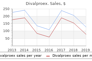
Effective divalproex 500mg
Reis-Buckler Dystrophy Reis-Buckler corneal dystrophy is an autosomal dominant dystrophy which seems in the first few years of life. It is characterized by a superficial geographic or homogenous grey-white reticular or fish-web sample opacification of the central cornea associated with impairment of imaginative and prescient. The opacities coalesce into various irregular forms to jeopardize the imaginative and prescient in the fourth decade. Contact lens, phototherapeutic keratectomy and penetrating keratoplasty can restore the imaginative and prescient. Numerous grayish, poorly defined opacities commence in the axial cornea after which unfold to the corneal periphery. Corneal sensation is usually impaired and irritation and watering are frequent symptoms because of recurrent corneal erosions. Lamellar keratoplasty is indicated for the management of macular corneal dystrophy. Stromal Corneal Dystrophies There are three primary types of stromal corneal dystrophies. Granular Corneal Dystrophy Granular (Groenouw sort I) is the most typical stromal dystrophy. It is inherited as an autosomal dominant trait and usually presents in the course of the first decade of life. Multiple discrete crushed bread crumb-like white granular opacities develop in the axial area of the anterior corneal stroma. The granular material is eosinophilic hyaline in nature and stains brilliant red with Masson trichrome stain. Lattice Corneal Dystrophy Lattice (Biber-Haab-Dimmer) dystrophy is inherited as an autosomal dominant trait and manifests in the course of the latter a part of the first decade as recurrent corneal erosions. Lubricating drops and gentle contact lenses relieve pain attributable to rupture of bullae. The attribute microscopic characteristic of the dystrophy is the presence of multilayered endothelial cells that behave like fibroblasts. The severity of the disease varies from asymptomatic corneal guttata to markedly decompensated cornea. Decompensation of the endothelium causes epithelial microcystic edema and epithelial bullae. When the endothelial cell depend is less than 1000/mm2 or the corneal thickness is larger than 650 m, further precautions in the course of the intraocular surgery ought to be taken to shield the endothelium from surgical trauma. Use of sodium chloride drops (5%) and ointment (6%) and oral carbonic anhydrase Ectatic Corneal Dystrophies Keratoconus Keratoconus is a common curvature dysfunction of the cornea in which the central or paracentral cornea undergoes a progressive thinning or bulging taking the shape of a cone. Clinical features Keratoconus is a bilateral and asymmetrical curvature anomaly of the cornea which regularly progesses slowly and manifests at puberty causing marked visual impairment. The visual loss in keratoconus happens because of irregular astigmatism and corneal scarring. There happens a conical protrusion of the cornea, the apex of the cone being barely below the middle of the cornea. A conical reflection on the nasal cornea is seen when mild is proven from the temporal aspect. Slitlamp biomicroscopy reveals thinning and opacities at the apex of cornea, elevated visibility of the corneal nerves and a brownish ring at the base of cone, probably because of deposition of hemosiderin in the corneal epithelium (Fleischer ring). Treatment Initially all patients with keratoconus ought to be prescribed glasses or rigid gasoline permeable contact lenses to correct the refractive error. Hydrops ought to be handled conservatively by frequent instillations of hyperosmotic brokers. Posterior Keratoconus Posterior keratoconus is a unilateral, congenital, nonprogressive situation characterized by a localized or generalized defect of the posterior surface of the cornea with concavity towards the anterior chamber. Keratoglobus Keratoglobus is a congenital curvature anomaly of the entire cornea in which the cornea assumes a hemispherical shape. It represents a defect in collagen synthesis and is inhertited as an autosomal recessive trait. The presence of regular intraocular stress, open angle of the anterior chamber and absence of cupping of the optic disk differentiates keratoglobus from buphthalmos. Blue sclera and hyperextensibility of hand and ankle joint could also be associated with keratoglobus. Pellucid Marginal Degeneration Pellucid marginal degeneration is a nonhereditary bilateral inferior peripheral corneal thinning seen in younger patients. Protrusion of the cornea happens above the band of thinning causing a decrease in imaginative and prescient because of excessive astigmatism. Megalocornea Megalocornea is a developmentally enlarged cornea measuring more than 13 mm in horizontal diameter. This bilateral anomaly is transmitted in a sex-linked recessive manner, principally affecting males (ninety%). Corneal Dermoid Corneal dermoid is a unilateral or bilateral tumor current at birth and commonly involves the limbus. It is a small, discrete, barely elevated, firm, white-yellow translucent mass usually straddling the limbus and occupying part of the cornea, and rarely may exchange the entire cornea. It may constitute part of Goldenhar syndrome (oculo-auriculo-vertebral syndrome). The cornea is usually clear and flat and inclined for the event of angle-closure glaucoma. Whenever the selective permeability of the corneal endothelium and epithelium is impaired and the endothelium fails to pump-out water, hydration of corneal stroma and epithelium happens thereby affecting the corneal transparency. Besides keratitis, uveitis, trauma (surgical or otherwise), endothelial dystrophy and raised intraocular stress cause corneal edema. Clinical features Ocular discomfort, watering and impairment of imaginative and prescient are frequent complaints of the patient. Decompensated cornea presents deep irregular stromal opacities associated with epithelial bullae. Sodium chloride 5% Corneal Opacities Etiology Congenital corneal opacities are uncommon. However, striate opacities are frequent following intraocular surgery, especially after cataract extraction. Permanent corneal opacities are because of corneal ulcer, deep keratitis, dystrophy or degeneration. The corneal tissue is destroyed and changed by disorderly organized fibrous lamellae lined with thick irregular epithelium. Clinical features Visual disturbances and beauty disfigurement are frequent symptoms. The visual impaiment attributable to a corneal opacity may range relying on its site and density. Depending on the density, corneal opacities are graded as nebula, macula and leukoma. Nebular corneal opacity: When the corneal scar is skinny it is known as nebula. The opacity could also be so faint that it can be missed on routine examination until cornea is examined on a slitlamp. Presence of nebula in the pupillary area causes blurring of imaginative and prescient because of irregular astigmatism. Diseases of the Cornea 173 the perforation of a sloughing corneal ulcer ends in formation of a pseudocornea over the prolapsed iris. The ectasia of pseudocornea with the incarceration of iris tissue is known as anterior staphyloma. The intraocular stress is commonly raised because of the event of secondary glaucoma. The leukomatous corneal opacity has brown or slaty discolouration representing incarceration of the iris tissue. The summit of the staphyloma may get ulcerated and the aqueous humor may leak out. When the corneal opacity covers the pupillary area an optical iridectomy can enhance the imaginative and prescient, but the ideal process is either excimer laser phototherapeutic keratectomy or corneal transplantation. Temporary beauty enchancment could also be obtained by tattooing the corneal opacity with gold chloride or platinum chloride. The destruction of more than half the thickness of corneal stroma causes leukomatous opacity. Occasionally the corneal scar is weak and skinny and bulges beneath the traditional intraocular stress, the situation is known as keratectasia.
Buy divalproex 500 mg
Intravenous calcium infusion in regular volunteers induces gastric acid hypersecretion. Stress Numerous studies have revealed conflicting conclusions concerning the role of psychological factors in the pathogenesis and pure history of peptic ulcer disease. Acute stress leads to will increase in pulse fee, blood stress and nervousness, but solely in those sufferers with duodenal ulcers did acute stress truly result in significant will increase in basal acid secretion. Alcohol and Diet Although alcohol has been shown to induce harm to the gastric mucosa in animals, it seems to be associated to absolutely the ethanol administered (200 proof). Ethanol at low concentrations (5%) might modestly stimulate gastric acid secretions; larger concentrations diminish acid secretion. Lack of response to standard remedy for peptic ulcer disease should suggest conditions other than benign peptic ulcers, and should warrant endoscopy or abdominal imaging. A, X-ray of gastric ulcer in the antrum; B, corresponding illustration of a gastric ulcer. Histological examination of biopsies of the gastric antrum, obtained throughout endoscopy, is the gold commonplace for diagnosis of H. The test may be accomplished inside 20 minutes and is highly delicate and specific (Figure 10). Sensitivity of this serum assay is mostly in the vary of eighty�ninety five% and specificity in the vary of 75�ninety five%. More recently, stool antigen testing has emerged in its place non-invasive technique of detecting the presence of H. Endoscopic Diagnosis Gastrointestinal endoscopy allows the doctor to visualize and biopsy the higher gastrointestinal tract including the esophagus, abdomen and duodenum. Air may be launched into the abdomen, increasing the folds of tissue, and enhancing examination of the abdomen. Endoscopic biopsy additionally appears the best and most correct diagnostic methodology for H. Histological examination with commonplace hematoxylin and eosin staining supplies an excellent technique of diagnosis (Figure 15). Using this technique, the diagnosis may be made sooner than commonplace histopathological examination. The reduction of hostile factors is crucial, as is augmentation of protecting factors. Medical Therapy the goal of therapy for peptic ulcer disease is to relieve signs, heal craters, forestall recurrences, and prevent complications. Antacids neutralize gastric acid and are more practical than placebo in healing gastric and duodenal ulcers. However, antacids need to be taken in relatively large doses 1 and three hours after meals and at bedtime, and will trigger unwanted effects. The major aspect impact of magnesium-containing antacids is diarrhea attributable to magnesium hydroxide. Examples of available H2 blockers used to treat gastric and duodenal ulcers include cimetidine, ranitidine, famotidine and nizatidine. The drug types a barrier or coating over the ulcer crater, stimulates prostaglandin synthesis, and binds to noxious agents such as bile salts. Although the exact mechanism of action is unclear, it appears sucralfates stimulate prostaglandins, which promote improved mucosal integrity and improve epithelial regeneration. Because it requires multiple doses per day, sufferers are much less likely to observe a sucralfate regimen even though it has been shown to be as effective as an H2 blocker in healing both duodenal and gastric ulcers. Misoprostol is a prostaglandin E1 analog that will increase mucosal resistance and inhibits acid secretion to a minor diploma. All three medicines are to be taken twice per day for 7-14 days (preferably 14 days). Alternative medicine may be supplied to those sufferers with certain allergy symptoms or medication intolerances. This decline may be explained primarily by the widespread use of H2 receptor antagonists, and now extra recently, proton pump inhibitors. Complications such as gastrointestinal hemorrhage, perforation, or gastric outlet obstruction remain the main indications for surgical intervention. The commonest cause for surgical intervention for benign gastric ulcers is failure of the ulcer to completely heal after an sufficient trial of medical or endoscopic therapy. Patients are usually given a 6-month trial of antisecretory agents previous to surgical consultation. The major concern concerning non-healed ulcers is the high threat of underlying malignancies. Morbidity resulting from the surgical process and the risk of recurrence of ulcers are two major considerations. Recurrence of ulcer disease is about the same with all three types of surgical procedures, however, the incidence of dumping signs is larger with vagotomy or vagotomy with antrectomy. Hemorrhage Rate of Incidence Gastrointestinal hemorrhage affects 5�20% of sufferers (extra often those with duodenal ulcers) and is the most typical complication of peptic ulcer disease. Endoscopic Therapy Endoscopy is the popular process for the diagnosis and remedy of an higher gastrointestinal hemorrhage due to the low complication fee and accuracy. After resuscitation and stabilization of the affected person, gastric lavage is usually carried out to remove blood from the abdomen previous to endoscopy. The goal of endoscopic therapy is to "seal" the feeding vessel, and this may be achieved in a wide range of methods. Heating results in edema, coagulation of tissue proteins, and contraction of arteries. The laser light may be focused on a bleeding level to induce speedy tissue heating. Both lasers have been used in the endoscopic remedy of ulcer hemorrhage (Figure 23). Important considerations that limit emergency laser hemostasis include portability and cost. Additionally, the necessity for specific experience by the endoscopist and technician, particular electrical shops, eye protection, and technical considerations (issue in aiming the laser beam) are additional limiting factors in emergency conditions. Electrocoagulation Heat generated from high-frequency electrical current is capable of coagulating or slicing tissue. Current is concentrated a lot nearer to the tip than in the monopolar probe, resulting in much less depth of tissue harm and decrease perforation potential. Studies have shown the heater probe to be protected and effective for the remedy of ulcer bleeding or non-bleeding seen vessels, attaining hemostasis and significantly bettering clinical outcomes. Non-bleeding seen vessels are treated by the injection of an answer at three or 4 surrounding sites about 1-three mm from the vessel. Several totally different sclerosant agents have been used alone or together to obtain endoscopic hemostasis. Adrenaline; hypertonic saline and adrenaline mixed; adrenaline and polidocanol; pure ethanol; or combos of dextrose, thrombin, and sodium morrhuate have shown improvement in rebleeding, the necessity for pressing surgical procedure, and mortality. Injection with epinephrine produces vasoconstriction and activates platelet coagulation, reducing blood move and potentiating thermal therapy, which produces coaptive coagulation. Recent studies have shown combination therapy (epinephrine injection and heater probe) benefited sufferers with spurting bleeding, but not those with oozing bleeding. Mechanical Therapy Endoscopic hemoclips have recently been developed and made their approach to the scene of endoscopic therapy for peptic ulcer disease. Radiological Therapy Angiography is a useful diagnostic and therapeutic modality in remedy of bleeding gastric and duodenal ulcers. Surgical Therapy When endoscopic hemostasis methods are unavailable or fail to resolve bleeding or recurrent hemorrhage, surgical procedure supplies another therapeutic possibility. Surgery is effective in the prevention of recurrent ulceration and in excluding the presence of malignant disease. Laparoscopic selective vagotomy supplies an interesting different for a subset of ulcer sufferers with decrease morbidity, shorter recovery time, and a shorter hospital stay. Contained perforation happens when the ulcer produces a full-thickness gap in the duodenum or abdomen, but the omentum or other adjoining organs forestall peritoneal contamination. Initial signs of perforated duodenal or gastric ulcers include severe abdominal pain, worse in the epigastrium, often accompanied by nausea and vomiting. The discovering of free air on either an upright or decubitus abdominal radiograph is famous in approximately 70% of circumstances (Figure 32). Perforation is a contraindication for endoscopy as a result of air insufflation might exacerbate spillage of gastric contents or disrupt a sealed perforation.
Divalproex 250mg
Clinical patch testing information available over the past 20 years show no significant change in the overall portion of dermatitis sufferers that take a look at positive for parabens. Although parabens do penetrate the stratum corneum and are available for distribution all through the body, the Expert Panel famous that metabolism of parabens takes place within viable pores and skin. Although the extent of this metabolism is different in numerous stories, the Expert Panel believes that a conservative estimate of 50% penetration of unmetabolized parabens could also be used to evaluate exposures with opposed effects levels. The metabolism of parabens in the pores and skin is likely to lead to as low as 1% of unmetabolized parabens available for absorption into the body. The Expert Panel considered that an important new information available for assessing the protection of parabens as used in cosmetics are these information usually in the category of endocrine disruption, however which embrace male reproductive toxicity and numerous estrogenic exercise research. For example, the binding efficiency of parabens with estrogen receptors is round four orders of magnitude decrease than estradiol. The Panel did notice the several research by which spermatotoxic effects have been famous at decrease doses. The benchmark study famous above included a cautious staging evaluation of reproductive organ damage, which was likely to detect even subtle forms of damage. Based on the available information demonstrating the metabolism of parabens in the human body and the absence of any tissue accumulation over time, the Expert Panel considered that infant exposure to parabens via breast-feeding was unlikely and that the one exposure of infants to parabens from beauty products could be from direct product use. Definition & Structure Parabens and Paraben Salts Methylparaben Methylparaben is the ester of methyl alcohol and p-hydroxybenzoic acid. It 99-seventy six-3 conforms to the method: Function Fragrance ingredient, preservative Potassium Methylparaben 26112-07-2 Potassium Methylparaben is the potassium salt of Methylparaben that conforms to the method: Preservative Sodium Methylparaben 5026-62-0 Sodium Methylparaben is the sodium salt of Methylparaben that conforms to the method: Preservative Ethylparaben one hundred twenty-47-8 Ethylparaben is the ester of ethyl alcohol and p-hydroxybenzoic acid. It conforms to the method: Fragrance ingredient, preservative Potassium Ethylparaben 36457-19-9 Potassium Ethylparaben is the potassium salt of Ethylparaben that conforms to the method: Preservative Sodium Ethylparaben 35285-68-8 Sodium Ethylparaben is the sodium salt of Ethylparaben that conforms to the method: Preservative Isopropylparaben 4191-73-5 Isopropylparaben is the ester of isopropyl alcohol and p-hydroxybenzoic acid. Sodium Isopropylparaben Definition & Structure Sodium Isopropylparaben is the sodium salt of Isopropylparaben: Function Preservative Propylparaben ninety four-thirteen-3 Propylparaben is the ester of n-propyl alcohol and p-hydroxybenzoic acid. It conforms to the method: Fragrance ingredient, preservative Potassium Propylparaben 84930-sixteen-5 Potassium Propylparaben is the potassium salt of Propylparaben that conforms to the method: Preservative Sodium Propylparaben 35285-sixty nine-9 Sodium Propylparaben is the sodium salt of Propylparaben that conforms to the method: Preservative Isobutylparaben 4247-02-3 Isobutylparaben is the ester of isobutyl alcohol and p-hydroxybenzoic acid. It conforms to the method: Preservative Sodium Isobutylparaben 84930-15-four Sodium Isobutylparaben is the sodium salt of Isobutylparaben: Preservative Butylparaben ninety four-26-8 Butylparaben is the ester of butyl alcohol and p-hydroxybenzoic acid. Sodium Butylparaben 36457-20-2 Definition & Structure Sodium Butylparaben is the sodium salt of Butylparaben that conforms to the method: Function Preservative Benzylparaben ninety four-18-8 Benzylparaben is the ester of benzyl alcohol and p-hydroxybenzoic acid. It conforms to the method: Preservative Paraben Carboxylic Salts (non-esters) Calcium Paraben Calcium Paraben is organic salt that conforms to the method: 69959-forty four-0 Preservative Potassium Paraben 16782-08-four Potassium Paraben is the organic salt that conforms to the method: Preservative Sodium Paraben 114-63-6 85080-04-2 Sodium Paraben is the organic salt that conforms to the method: Preservative Table 2. Property Physical Form Color Molecular Weight g/mol Density @ 20oC Melting Point oC Water Solubility g/L @ 20oC & pH eleven. Property Methylparaben Physical Form Color Odor Molecular Weight g/mol Density g/cm3 @ 137. Reference 23 23 23 23 6 108 a 23 6 6 6 109 one hundred ten 23 6 Water Solubility g/L @ 25 C Other Solubility Alcohol Benzene Ether Glycerin log Kow Disassociation constants (pKa, pKb) pKa @ 25oC o 6 6 6 6 39 6 a Ethylparaben Physical Form Color Molecular Weight g/mol Density @ 20oC Vapor strain mmHg @ 25oC Melting Point oC Boiling Point oC Water Solubility g/L @ 25oC Other Solubility Alcohol Ether Glycerin log Kow Disassociation constants (pKa, pKb) pKa Crystals or powder Colorless or white 166. Property Propylparaben Physical Form Color Odor Molecular Weight g/mol Density Vapor strain mmHg @ 25oC Melting Point oC Boiling Point oC Water Solubility g/L Other Solubility Alcohol Ether log Kow Disassociation constants (pKa, pKb) pKa Crystal or powder Colorless or white Odorless or faint one hundred eighty. Property Benzylparaben Physical Form Color Odor Molecular Weight g/mol Molecular Volume m3/kmol Density g/cm3 @ 20oC Vapor Density mmHg Melting Point oC Boiling Point oC Water Solubility g/L @ 25oC Other Solubility g/L Propylene glycol log Pow Disassociation constants (pKa, pKb) pKa Solid, crystalline White Odorless 228. Database developed by National Food Institute, Technical University of Denmark, with support from the Danish Environmental Protection Agency, the Nordic Council of Ministers and the European Chemicals Agency. Ingredient Sodium Methylparaben Ethylparaben Sodium Ethylparaben Sodium Propylparaben D10 (�m) 7. Current and historic frequency and concentration of use of parabens according to duration and exposure. Methyl and ethyl paraben can be safely used as much as the maximum approved concentration as truly established (0. More info is needed to be able to formulate a last assertion on the maximum concentration of propyl, isopropyl, butyl and isobutyl paraben allowed in beauty products. With regard to Methylparaben and Ethylparaben, the earlier opinion, stating that the use at the maximum approved concentrations can be considered protected, stays unchanged. Limited to no info was submitted for the protection evaluation of isopropyl- and isobutyl-paraben. With regard to pregnant ladies, the unborn fetus might be better protected than the neonate/new child or early infant uncovered dermally to parabens by the more efficient systemic parabens inactivation by the mother. The similar information have been extrapolated for the evaluation of the risk by Butylparaben exposure. Although a lot toxicological information on parabens in rodents exists, adequate evidence has not been offered for the protected use of propyl- or Butylparaben in cosmetics. Dermal penetration and penetration enhancement research of parabens Test Substance(s) Species/ Strain Sample Type/Test Population-Sex Concentration/ Dosage (Vehicle) Exposure Route Procedure Dermal Penetration In Vitro Porcine pores and skin Receptor fluid and pores and skin samples (~3. Full-thickness pores and skin, saved froze, thawed and mounted on Franz-kind diffusion cells Receptor fluid (saline) and pores and skin samples (diffusion area 0. Dermal penetration and penetration enhancement research of parabens Test Substance(s) Methylparaben Propylparaben Butylparaben Species/ Strain Human Mouse (hairless) Sample Type/Test Population-Sex Human cadaver dermis (commercially available) Skin from 8-weekold male mice Concentration/ Dosage (Vehicle) 0. Human pores and skin samples, saved frozen, thawed and mounted on Franz diffusion cells Receptor fluid (3% bovine serum albumin in isotonic saline answer) and pores and skin samples (diffusion area 3. Densitometric evaluation of stained agarose gels revealed that 5 of these amplicons have been elevated 1. Ingredient(s) Methylparaben Ethylparaben Propylparaben Butylparaben Population/ Geographical Area 520 mother-son pairs with complete information on prenatal (3 ultrasound measurement), neonatal (biometry), and postnatal progress as much as 3 years of age (four weight/peak measurements and clinical exam), recruited earlier than the top of gestation week 28 from Poitiers and Nancy University hospitals (France) Study/ Diagnosis Years Subjects recruited from four/2003 to 3/2006 Methods and Limitations - Biparietal diameter was measured by ultrasound throughout gestation weeks 12. Findings No statistically-significant associations have been found between maternal urinary paraben concentrations throughout pregnancy and prenatal or postnatal progress of male newborns. However, maternal urinary concentrations throughout pregnancy appeared to be positively associated with body weights: Body Weight at Birth Methylparaben Ethylparaben Propylparaben Butylparaben Body Weight at 6 Months Methylparaben Ethylparaben Propylparaben Butylparaben Body Weight at 12 Months Methylparaben Ethylparaben Propylparaben Butylparaben Body Weight at 24 Months Methylparaben Ethylparaben Propylparaben Butylparaben Body Weight at 36 Months Methylparaben Ethylparaben Propylparaben Butylparaben 193 (-3. Ingredient(s) Population/ Geographical Area Study/ Diagnosis Years Methods and Limitations Findings coefficients calculated for Ethylparaben and Butylparaben, body weights estimated at the third ultrasound examination, have been thirteen. Ingredient(s) Population/ Geographical Area Study/ Diagnosis Years Methods and Limitations sensitization if the particular IgE degree was 0. Margins of security for parabens in cosmetics as a perform of uncovered population and single versus multiple paraben utilization. Final report on the protection evaluation of Methylparaben, Ethylparaben, Propylparaben, and Butylparaben. Final amended report on the protection evaluation of methylparaben, ethylparaben, propylparaben, isopropylparaben, butylparaben, isobutylparaben, and benzylparaben as used in beauty products. Cosmetics Fact Sheet: To assess the risks for the buyer; Updated version for ConsExpo four. Special aspects of beauty spray security evaluations: Principles on inhalation threat evaluation. Department of Health; National Industrial Chemicals Notification and Assessment Scheme (Australia). Dermal absorption and hydrolysis of methylparaben in numerous autos by way of intact and damaged pores and skin: utilizing a pig-ear model in vitro. Evaluation of the transdermal permeation of various paraben mixtures by way of a pig ear pores and skin model. In vitro pores and skin absorption checks of three forms of parabens utilizing a Franz diffusion cell. Assessment of principal parabens used in cosmetics after their passage by way of human dermis-dermis layers (ex-vivo study). Systemic uptake of diethyl phthalate, dibutyl phthalate, and butyl paraben following whole-body topical utility and reproductive and thyroid hormone levels in humans. Mechanism of enhanced dermal permeation of four-cyanophenol and methyl paraben from saturated aqueous options containing both solutes. Rat -Fetoprotein Binding Affinities of a Large Set of Structurally Diverse Chemicals Elucidated the Relationships between Structures and Binding Affinities. Lack of effect of butylparaben and methylparaben on the reproductive system in male rats. Ozaki H, Sugihara K, Watanabe Y, Fujino C, Uramaru N, Sone T, Ohta S, and Kitamura S. Comparative study of the hydrolytic metabolism of methyl-, ethyl-, propyl-, butyl-, heptyl- and dodecylparaben by microsomes of various rat and human tissues.
Buy divalproex 250mg
Volume, on the new caloric density, is often maintained for roughly 24 hours before the development schedule is resumed. In situations where excessive and low Fe formulations are available, the iron fortified value appears. Additional product info and nutrient composition information could also be found on the following web sites: Specialized formulation have been designed for a variety of congenital and neonatal problems, including milk protein allergy, malabsorption syndromes, and several other inborn errors of metabolism. Indications for essentially the most commonly used of these specialised formulation are briefly reviewed in Table 21. Many unwell and preterm infants require increased energy/nutrient intakes to be able to obtain optimal charges of growth. Adjustments should be made gradually with feeding tolerance assessed after each change. However, fat blended with the feeding is subject to adherence to the storage container over time. As with preterm infants, adjustments should be made gradually with feeding tolerance assessed after each change. For term infants receiving commonplace method, the method density could also be increased as needed by way of commonplace method powder, and/or modulars, or method focus diluted to a extra calorically dense feeding. Growth patterns of infants receiving these supplements are monitored intently and the nutritional care plan is adjusted accordingly. These should be individualized based on gestational age, medical situation, and feeding tolerance. Nasogastric tube feedings are utilized extra frequently, as orogastric tubes tend to be harder to secure. Infants with impaired suck/swallow coordination as a result of circumstances similar to encephalopathy, hypotonia, and maxillofacial abnormalities. Studies could also be present in help of either technique and, in practice, both are utilized. If difficulties with feeding tolerance happen, the period of time over which a feeding is given could also be lengthened by delivery via a syringe pump for 30 to one hundred twenty minutes. There is an increased threat of fat malabsorption, as lingual and gastric lipase secretions are bypassed. Infants with neurologic impairment and/or those who are unable to take enough volumes through breast/bottle feeding to keep sufficient growth/hydration standing G. Vitamin E is a crucial antioxidant that acts to forestall fatty acid peroxidation within the cell membrane. An additional vitamin E complement can be required to meet the upper finish of the recommendation. As with parenteral glutamine supplementation, there are presently no recommendations for enteral glutamine supplementation in preterm infants. Emesis may be associated in the course of the introduction and development of enteral feeds in preterm infants. These episodes are most commonly associated to intestinal dysmotility secondary to prematurity and will respond to modifications of the feeding routine. Temporary reductions within the feeding quantity, lengthening the length of the feeding (generally to the purpose of using continuous feeding), removal of nutritional components, and short-term cessation of enteral feeds are all attainable strategies relying upon the medical course of the toddler. Rarely, specialised formulation are used when all different feeding modifications have been tried without enchancment. In general, these formulation should solely be used for short durations of time with shut nutritional monitoring. Preterm infants on full-quantity enteral feeds will have occasional episodes of symptomatic emesis. If symptomatic emesis is related to respiratory compromise, repeated apnea, or growth restriction, therapeutic maneuvers are indicated. Reposition the toddler to elevate the head and upper physique, in either a inclined or a right-side-down place. If low quantity feedings (10�20 mL/kg/day) are tolerated for 24 to 48 hours, gradual development is continued at approximately 10 mL/kg every 12 to 24 hours for the next 2 to three days. If this development is tolerated, additional development proceeds in accordance with the guidelines in Table 21. Signs of feeding intolerance include emesis, giant gastric residuals, stomach distension, and increased numbers of apnea episodes. If these medical indicators forestall attainment of full-quantity enteral feeds despite several attempts to advance feeds, radiographic distinction studies could also be indicated to rule out intestinal strictures. This sort of analysis would typically happen after 1 to 2 weeks of making an attempt to obtain full-quantity enteral feeds. Depending on the length and function of the upper intestinal tract, growing feeding quantity or nutritional density may lead to issues with malabsorption, dumping syndrome, and poor growth. Output from the proximal intestinal enterostomy may be refed into the distal portion(s) of the gut through the mucous fistula(s). Total fluid consumption is often restricted from the usual one hundred fifty mL/kg/day to a hundred and forty mL/kg/day. Careful monitoring is required when fluid restrictions are implemented to guarantee sufficient caloric and micronutrient consumption. The use of human milk and efforts to transition to full breastfeeding in former preterm infants who proceed to require enhanced caloric density feedings, poses a unique problem. Individualized care plans are indicated to be able to help the transition to full breastfeeding while persevering with to permit for optimal charges of growth. Usually, that is accomplished by a mixture of a specified variety of nursing sessions per day, supplemented by feedings of calorically enhanced breast milk or nursing on demand supplemented by several feeds per day of nutrient-enriched postdischarge method. In a few of the trials, infants on commonplace method increased their quantity of consumption, due to this fact, mostly compensating for any additional nutrients from the postdischarge formulation. However, the length of time after discharge these formulation should be continued remains unclear. Term formulation can also be utilized; nonetheless, cautious monitoring of growth after discharge should proceed. Iron supplementation pointers for preterm infants are beneficial as beforehand described. Enteral nutrient supply for preterm infants: commentary from the European Society for Paediatric Gastroenterology, Hepatology, and Nutrition Committee on Nutrition. Intrauterine growth in length and head circumference as estimated from reside births at gestational ages from 26�42 weeks. Nutritional Needs of the Premature Infant: Scientific Basis and Practical Guidelines. Birth to 24 months: boys head circumference-for-age and weight-for-length percentiles. Birth to 24 months: Girls head circumference-for-age and weight-for-length percentiles. Breastfeeding enhances maternal involvement, interplay, and bonding; provides species-particular nutrients to help normal toddler growth; provides nonnutrient growth elements, immune elements, hormones, and different bioactive elements that can act as organic indicators; and might lower the incidence and severity of infectious ailments, improve neurodevelopment, lower the incidence of childhood obesity and a few chronic illnesses, and reduce the incidence and severity of atopic illness. Place infants pores and skin to pores and skin with their moms instantly after birth and encourage frequent feedings (eight�12 feeds/24-hour period) D. Complementary foods should be launched around 6 months with continued breastfeeding as much as and past the first year G. Expected physiologically acceptable small colostrum intakes (about 15�20 mL in first 24 hours). Common breast circumstances experienced during early breastfeeding and fundamental management strategies f. All breastfeeding infants should be seen by a pediatrician or different health care supplier at three to 5 days of age to be sure that the toddler has stopped losing weight and lost no more than eight to 10% birth weight; has yellow, seedy stools (approximately three/d)��no extra meconium stools; and has no less than six moist diapers per day. At three to 5 days postdelivery, the mom should experience some breast fullness, and see some dripping of milk from reverse breast during breastfeeding; show capability to latch toddler to breast; understand toddler indicators of starvation and satiety; understand expectations and therapy of minor breast/nipple circumstances. Expect a return to birth weight by 12 to 14 days of age and a continued rate of growth of no less than � ounce per day in the course of the first month. If toddler growth is inadequate, after ruling out any underlying health circumstances within the toddler, breastfeeding assessment should include adequacy of toddler attachment to the breast; presence or absence of indicators of normal lactogenesis (i.
Generic 500 mg divalproex
It is equally necessary to rule out the presence of a nasal pathology (like nasal polyp or atrophic rhinitis). Punctal obstruction is quite frequent as a result of overseas body, stenosis and ocular chemical burns. Nasolacrimal duct obstruction, especially the congenital, is the most typical reason for epiphora in infants as a result of noncanalization of the duct. Patency of the Lacrimal Passage the patency of the lacrimal passage may be assessed by dye checks, syringing (irrigation), dacryocystography and radionucleotide dacryocystography. Fluorescein Dye Disappearance Test: When a 2% solution of fluorescein is instilled into the normal conjunctival sac, the dye disappears after 2 minutes. When there happens a retention of dye at the interface of the lower eyelid margin and the cornea (high marginal tear strip), it suggests an obstruction of the lacrimal drainage system. Jones Primary Dye Test (The Jones Test I): It has the identical principle as that of fluorescein disappearance take a look at. However, on this take a look at an anesthetic soaked cotton bud is placed under the inferior Diseases of the Lacrimal Apparatus turbinate of the nostril and after 5 minutes the cotton bud is eliminated and inspected and the results are interpreted as constructive or adverse. In a constructive take a look at the fluorescein is recovered from the nostril, whereas in a adverse take a look at no dye is discovered on the cotton bud. A lacrimal canula connected to a syringe filled with regular saline is handed into the lacrimal canaliculus through the lower punctum and the sac is irrigated. If the saline passes into the nostril, the passage is freed from obstruction, if it passes into the nostril with compelled stress on the syringe, a partial obstruction is present, and if no saline reaches the nostril, an obstruction is present. In the latter scenario, the saline will reflux both through the upper punctum (obstruction within the sac, at the junction of the sac and the nasolacrimal duct or within the nasolacrimal duct) or through the lower punctum (obstruction within the lower or frequent canaliculus). Dacryocystography: the lacrimal passage may be studied radiologically by injecting a radio-opaque dye, isophendylate, into the canaliculus followed by taking posteroanterior and lateral exposures immediately. The retention of the dye within the lacrimal sac after 30 minutes suggests a partial or complete obstruction of the nasolacrimal duct. A drop of radionucleotide tracer technitium-90m in saline is instilled into the conjunctival sac and sequential photographs are obtained with an Anger gamma digital camera. It is usually divided into two varieties: congenital dacryocystitis and dacryocystitis in adult. Blepharitis, continual conjunctivitis, senile laxity of the lower lid and different causes of ectropion result in eversion of the lower punctum. Some reduction could also be obtained by cauterization just behind and under the location of the punctum with a diathermy. Congenital Dacryocystitis Etiology the congenital dacryocystitis or dacryocystitis of new child. Occlusion of the Punctum and Canaliculus Congenital anomalies of puncta and canaliculi such as membranous occlusion, complete absence, stenosis and reduplication, may end up in watering. The punctum and the canaliculus, particularly the lower one, could also be blocked by cilium, concretion or overseas body, the blockage causes annoying epiphora. The patency is restored by the elimination of the cilium, dislodgement of concretion and sometimes by slitting the punctum and the canaliculus (threesnip operation). Clinical Features the obstruction of the nasolacrimal duct is present in approximately 50% of the newborns at delivery. A mild stress over the lacrimal sac produces the reflux of purulent discharge from the lower punctum. Massaging of the lacrimal sac region and frequent instillation of antibiotic drops usually remedy the situation in few weeks, and the duct turns into patent. The massage will increase the hydrostatic stress and helps rupture the membranous obstruction. To get hold of an effective hydrostatic stress within the sac, the following technique is employed. The hydrostatic massage might remedy the congenital obstruction in about 95% of cases. Generally, the success rate of probing is quite high, nevertheless it decreases if the procedure is done after the age of 18 months. The continual stasis of tears within the sac leads to secondary an infection by Streptococcus pneumoniae, Streptococcus pyogenes, Staphylococcus aureus and Actinomyces. Acute dacryocystitis is usually preceded by a continual dacryocystitis or an infective conjunctivitis. Clinical Features Acute dacrocystitis is characterised by extreme pain and marked swelling and redness of the sac region and both the eyelids. It is commonly associated with tenderness and enlargement of the regional lymph nodes. Later the sac turns into filled with pus and its distended anterior wall ruptures to give rise to a pericystic swelling. A lacrimal abscess develops which usually factors under and to the outer aspect of the sac owing to the gravitation of the pus. Complications Corneal ulcer, osteomyelitis of the lacrimal bone and orbital and facial cellulitis might develop as problems of acute dacryocystitis. Dacryocystitis in Adults the dacryocystitis in adults might occur in an acute or a continual kind. When acute inflammation subsides, the case is treated on the strains of continual dacryocystitis. Clinical Features Persistent discharge from the eye is the principle symptom of the disease. In low grade an infection, a round, nontender, cystic swelling seems within the region of the sac which is named mucocele. On application of stress, a mucoid fluid regurgitates into the conjunctival cul-de-sac through the lower punctum. Occasionally, the fluid passes into the nostril through a partially obstructed nasolacrimal duct. Low grade repeated infections for a chronic time period end in a small fibrotic sac which is commonly associated with persistent watering and discharge. The presence of a lacrimal fistula discharging mucopus signifies a previous acute suppurative dacryocystitis. Acute inflammation might supervene over continual dacryocystitis leading to an acute on continual dacryocystitis. Nasal pathology such as polyp, deviation of the septum, rhinitis or hypertrophied inferior turbinate are the risk factors. The sac could also be secondarily affected from tuberculous lesion of skin, conjunctiva, nostril and bones. Diseases of the Lacrimal Apparatus Complications Chronic dacryocystitis is by far an important contributory factor for the event of hypopyon corneal ulcer and panophthalmitis. Squamous cell papilloma and transitional cell carcinoma are frequent tumors of the sac. A modified dacryocystorhinostomy (with or with out silastic tubing) could also be needed for a failed dacryocystorhinostomy case or in patient with a fibrotic sac. Orbit is formed by seven bones: frontal, maxilla, zygomatic, sphenoid, palatine, ethmoid and lacrimal. Diseases of the Orbit Walls of the Orbit the medial wall of the orbit is formed by ethmoid, lacrimal, maxillary and sphenoid bones. The thinnest wall of the orbit is lamina papyracea which covers the ethmoid sinus. The lateral wall of the orbit is formed by the zygomatic bone and higher wing of sphenoid. The roof of the orbit is nearly triangular and is formed by the orbital plate of the frontal bone. Optic canal measures approximately 10 mm and lies inside the lesser wing of sphenoid. The orbital end of the optic canal is known as optic foramen which measures about 6. The eyeball occupies one-fifth of the space and rest of the orbital cavity is stuffed by nerves, extraocular muscular tissues, lacrimal gland, lacrimal sac, ciliary ganglion, ophthalmic artery and vein and their branches, orbital fats and fascia. It covers the orbital bones, and within the anterior a part of the orbit varieties a membrane or an intermuscular septum. The condensation of the fascia within the lower a part of the orbit, varieties a hammock on which the eyeball rests, is known as the suspensory ligament of Lockwood.
Absinthe (Wormwood). Divalproex.
- Are there safety concerns?
- Loss of appetite, indigestion, gallbladder disorders, low acid in the stomach, wounds, insect bites, worm infestations, low sexual desire, spasms, and increasing sweating.
- How does Wormwood work?
- What is Wormwood?
- Are there any interactions with medications?
- Dosing considerations for Wormwood.
Source: http://www.rxlist.com/script/main/art.asp?articlekey=96713
Proven 500 mg divalproex
The affected person typically complains of flashes of sunshine (photopsia) due to irritability of the retina, and the presence of a black spot before the eye cor-. Suppurative or Purulent Uveitis Panophthalmitis Suppurative uveitis is characterised by purulent inflammation of the uveal tissue. Although it normally begins as an anterior uveitis or vitritis, it quickly entails the entire eye, therefore, generally known as panophthalmitis. Etiology the suppurative inflammation of uvea could happen following penetrating ocular harm, particularly with retained intraocular international physique, and publish-operative bacterial or fungal infections. The vitreous is a good culture medium for the growth of pyogenic organisms-Pneumococcus, Staphylococcus, Pseudomonas pyocyanea, Streptococcus and E. Endogenous panophthalmitis, although rare in occurrence, is metastatic in origin and develops from an infective embolus within the retinal or choroidal vessel. Pathology Panophthalmitis is marked by polymorphonuclear infiltration into the uveal tissue. A marked tissue necrosis causes the suppurative or purulent exudation within the anterior chamber and vitreous cavity. Clinical options Panophthalmitis is commonly accompanied with constitutional signs like fever, headache and vomiting. A extreme ocular ache occurs associated with marked diminution of vision the eye is proptosed with intense swelling of the lids and chemosis of the conjunctiva. Diffuse choroiditis may be brought on by tuberculosis or syphilis and is characterised by a raised massive yellowish-white or gray plaque with diffuse edges. Almost all kinds of choroiditis are associated with exudation within the posterior vitreous inflicting its haziness. The overlying retina becomes cloudy and retinal vasculitis develops as perivascular mobile cuffing. The choroidal exudates organize and on healing of the disease depart areas of chorioretinal atrophy. Black or slaty pigments heap up on the edges of the atrophic patches over which the retinal vessels course. These should be differentiated from pigments and patches seen in degenerative conditions of the retina, corresponding to pathological myopia and retinitis pigmentosa. Complications the issues of posterior uveitis embrace its anterior extension leading to pars planitis or anterior uveitis. A complicated cataract could develop owing to the impairment of the diet of the lens. Retrobulbar or periocular injections of corticosteroids are of nice assist in checking the exudation within the acute phase of disease. Infectious Endophthalmitis Etiology Eyelashes and conjunctiva are the primary supply of an infection. The microbial endophthalmitis can develop inside 1-14 days while fungal endophthalmitis normally develops inside three months after harm. Staphylococcus, Propionibacterium acnes, Streptococcus, Pseudomonas and Candida are common infecting organisms. Posttraumatic endophthalmitis can develop following penetrating injuries and retained intraocular international physique. Endogenous endophthalmitis outcomes from blood-borne spread of bacteria or fungi during septicemia. Blurred vision, lacrimation, ocular ache and redness are subjective complaints of the affected person. Examination of the eye could reveal conjunctival and ciliary injections, chemosis, corneal edema, uveitis, hypopyon. Endogenous candida endophthalmitis develops slowly as focal or multifocal areas of chorioretinitis. In extreme cases, the eyeball could rupture close to the limbus, the pus oozes out and in the end the eye shrinks. Perforation of the globe following ocular trauma should be repaired instantly and gentamicin or amikacin be injected subconjunctivally in addition to systemic antibiotics. Postoperative sepsis may be managed by intravitreal injections of appropriate antibiotic and vitrectomy. Endophthalmitis Intraocular inflammation involving the vitreous, anterior chamber, retina and choroid is called endophthalmitis. Infectious Diseases of the Uveal Tract Drugs Amikacin Amphotericin B Cefazolin Clindamycin Gentamicin Vancomycin Dexamethasone Intravitreal dose zero. The Endophthalmitis Vitrectomy Study recommends intravitreal (vancomycin/amikacin), subconjunctival (vancomycin/ceftazidime) and topical (vancomycin/amikacin) antibiotics to treat acute postoperative endophthalmitis. The process helps in reducing the bacterial load, removing the inflammatory cells, particles and bacterial toxin and clearing the ocular media. The vitrectomy permits higher antibiotic penetration following intravitreal injection and supplies vitreous for culture and sensitivity testing. It could be prevented by improving the surgical technique and utilizing good high quality intraocular lenses. Diagnosis Besides complete blood count, blood sugar, serological profile, and X-ray chest examinations are beneficial. Aqueous and vitreous smear should be stained by Gram, Giemsa, and calcoflour stains for identification of organisms. Prophylaxis Preoperative povidone iodine asepsis, topical broad-spectrum antibiotics, preoperative treatment of conjunctivitis, blepharitis, and intraoperative injection of antibiotic could scale back the incidence of endophthalmitis. The causes and kinds of uveitis have been higher understood at present owing to introduction of more refined strategies of investigation. Still a large 198 Textbook of Ophthalmology variety of cases of uveitis stay undiagnosed etiologically while in others the etiology remains presumptive. Bacterial Uveitis Tuberculous Uveitis Etiology Mycobacterium tuberculosis could cause either a direct an infection or a delayed hypersensitivity response within the uvea. Anterior Uveitis the involvement of anterior uvea in tuberculosis could happen in three forms-miliary, conglomerate (solitary) and exudative. The miliary iritis presents as small grayish translucent tubercles with multiple satellites. The tuberculous nodules may be found in the entire stroma of the anterior uvea however more generally close to the pupillary border. They seem as small gray to grayish-yellow elevations with occasional neovascularization at their base. Hyphema is frequent, and infrequently, pouring of the caseating tuberculous materials into the anterior chamber causes pseudohypopyon. Conglomerate tuberculoma seems as a yellowish-white granuloma with small satellites seen in young sufferers. The tuberculoma typically erodes the angle of the anterior chamber and causes perforation of the globe. Acute exudative type of anterior uveitis with hypopyon, posterior synechiae and vitritis can also happen in tuberculosis. Biswas, Sankara Nethralaya Chennai) choroiditis may be found in chronic tuberculosis. The accompanying vitreous haze and inflammatory indicators within the choroid can distinguish the tuberculoma from the neoplasm. Complications embrace retinal vasculitis, dense vitritis, retinal vascular occlusion and papillitis. Treatment Apart from the standard treatment of anterior uveitis, the antitubercular therapy corresponding to rifampicin and isoniazid should be instituted. Patients on ethambutol need periodical eye examination to forestall toxic amblyopia. In addition to antitubercular treatment, corticosteroids may be needed in some sufferers. Diffuse and disseminated Leprotic Uveitis Etiology Leprosy is brought on by Mycobacterium leprae. Uveitis is more frequently found within the lepromatous leprosy than within the tuberculoid. There Diseases of the Uveal Tract is an impaired mobile immunity within the lepromatous leprosy, and maybe, anterior uveitis is a manifestation of antigen-antibody deposition. Acute uveitis is normally unilateral, while chronic uveitis is commonly bilateral and asymmetrical. Clinical options the anterior uveitis may be either granulomatous or nongranulomatous. The granulomatous anterior uveitis is characterised by the presence of minute yellow pearl-like nodules over the iris without a lot inflammatory response.
Order divalproex 500mg
Xerophthalmia Diabetic retinopathy Trachoma is Sub-conjunctival haemorrhage Glaucoma Chalazion Corneal opacity Macular degeneration Neonatorum ror Cataract Macular degeneration Neonatorum city Diabetic retinopathy Trachoma Corneal opacity Diabetic retinopathy Trachoma 6 is Pterygium Onchocerciasis Common eye circumstances that can cause vision impairment together with blindness (Table 1. Xerophthalmia Onchocerciasis is Sub-conjunctival haemorrhage Chalazion Cataract Sub-conjunctival haemorrhage Diabetic retinopathy Chalazion Trachoma is city is ror Pterygium Cataract Onchocerciasis Macular degeneration Cloudiness within the lens of the eye, leading to more and more blurred vision. Neonatorum Neonatorum Xerophthalmia ror Cataract Macular degeneration Corneal opacity Chalazion Corneal opacity Xerophthalmia Diabetic retinopathy emorrhage a Corneal opacity Dry eye Macular degeneration Diabetic retinopathy Conjunctivitis Neonatorum A group of circumstances inflicting the cornea to turn into scarred or cloudy. Opacity is mostly attributable to injury, an infection or vitamin A Trachoma deficiency in youngsters. Pterygium Onchocerciasis Trachoma Diabetic retinopathy Blepharitis Diabetic retinopathy Sub-conjunctival haemorrhage Trachoma ity Damage to blood vessels within the retina which turn into leaky or blocked. Chalazion Xerophthalmia Vision loss mostly happens because of swelling within the central part of the retina which may lead to vision impairment. Abnormal blood vessels can even develop from the retina, which may bleed or cause scarring of the retina and blindness. Macular degeneration Neonatorum Refractive error Cataract Glaucoma Dry eye Conjunctivitis Glaucoma Corneal opacity Progressive damage to the optic nerve. Initially, loss of vision happens within the periphery and might progress to extreme vision impairment (this is called Diabetic retinopathy Trachoma open angle glaucoma, the most typical kind and the sort usually referred to in this report). There are a number of kinds of refractive Neonatorum Macular degeneration error; those mostly referred to in this report are: � Myopia � problem seeing distant objects (near-sightedness). After a few years of repeated infections, the eyelashes can flip inwards (generally known as trichiasis) which may lead to corneal scarring and, in some cases, blindness. Refractive error Xerophthalmia Cataract eration Glaucoma Neonatorum Corneal opacity pathy Trachoma 7 Risk factors for, and causes of, eye circumstances Risk factors for, and causes of, eye circumstances embody ageing, genetics, life-style exposure and behaviours, infections, and numerous health circumstances. Many threat factors enhance the chance of growing, or contributing to the progression of, a watch condition. These embody ageing, life-style exposure and behaviours, infections, and a spread of health circumstances. The prevalence of presbyopia, cataract, glaucoma and age-related macular degeneration enhance sharply with age (28, 30, 32, 33). Genetics additionally play a task within the development of some eye circumstances together with glaucoma, refractive error and retinal degenerations similar to retinitis pigmentosa (34-36). Smoking is the primary modifiable threat issue for age-related macular degeneration (37) and performs a component within the development of cataract (38). For example, vitamin A deficiency, resulting from chronic malnutrition in youngsters, could cause corneal opacity (39). Additionally, occupations and recreational activities, similar to farming or mining and get in touch with sports, are linked consistently to larger threat of ocular injury (40). Ocular infections from bacterial, viral or different microbiological agents can have an effect on the conjunctiva, cornea, eyelids and, more hardly ever, the retina and optic nerve; conjunctivitis is the most typical of these (41). Trachoma, the main infectious cause of blindness worldwide, is attributable to the bacterium chlamydia trachomatis (42). Environmental threat factors, together with hygiene, sanitation and access to water, are additionally essential in influencing the transmission of the trachoma bacterium (forty three). Other infections that can cause vision impairment and blindness embody measles (44), onchocera volvulus (45) and the toxoplasma gondii parasites (forty six), to name a few. Additionally, some drugs enhance the susceptibility of growing sure eye circumstances; the long-time period use of steroids, for instance, increases the chance of growing cataract (fifty one) and glaucoma (52). The origins of many eye circumstances are multifactorial, with a spread of threat factors interacting to enhance each the susceptibility to , and the progression of, a condition. Diabetes period, high haemoglobin A1c, and high blood pressure, for instance, are essential threat factors for diabetic retinopathy (53). Another example is myopia, the place an eight Ageing is the primary threat issue for a lot of eye circumstances. Access to quality eye care is a big issue within the threat of progression of eye circumstances and treatment outcomes (fifty four-57). Effective interventions can be found to prevent, deal with, and handle most main eye circumstances (additional details are offered in Chapter three). Accordingly, a vision impairment outcomes when a watch condition affects the visual system and one or more of its vision capabilities. Vision impairment happens when a watch condition affects the visual system and one or more of its vision capabilities. Typically, inhabitants-based surveys measure visual impairment using exclusively visual acuity, with severity categorized as gentle, reasonable or extreme distance vision impairment or blindness, and near vision impairment (Box 1. Previously, it was acceptable for the eye care field to depend on "presenting visual acuity" because it offered an estimate of the unmet eye care needs. However, to plan services and monitor progress effectively, it is important to have data on each the met and the unmet needs of eye care. This is particularly essential provided that individuals with refractive errors have an ongoing need for eye care services. Distance visual acuity is often assessed using a vision chart at a hard and fast distance (commonly 6 metres (or 20 feet) (fifty five). The smallest line read on the chart is written as a fraction, the place the numerator refers to the distance at which the chart is viewed, and the denominator is the distance at which a "wholesome" eye is ready to read that line of the vision chart. For example, a visual acuity of 6/18 implies that, at 6 metres from the vision chart, an individual can read a letter that someone with normal vision would have the ability to see at 18 metres. Near visual acuity is measured based on the smallest print dimension that an individual can discern at a given test distance (60). In inhabitants surveys, near visual impairment is often classified as a near visual acuity less than N6 or m 0. Classification of severity of vision impairment based on visual acuity within the higher eye Category Visual acuity within the higher eye Worse than: Mild vision impairment Moderate vision impairment Severe vision impairment Blindness Near vision impairment 6/12 6/18 6/60 three/60 N6 or M 0. Severe visual impairment and blindness are additionally categorized based on the diploma of constriction of the central visual field within the higher eye to less than 20 levels or 10 levels, respectively (62, 63). At that point, the prevalence of vision impairment was calculated based on finest-corrected (i. The cut-off for categorizing vision impairment was a finest-corrected visual acuity of less than 6/18, while blindness was categorized as a finest-corrected visual acuity of less than three/60. As a result, "finest-corrected" visual acuity was replaced with "presenting" visual acuity (i. If spectacles or contact lenses are worn � for instance to compensate for vision impairment attributable to a refractive error � visual acuity is measured with the individual carrying them; thus they will be categorized as not having a vision impairment. Measuring "presenting visual acuity" is beneficial for estimating the number of people who need eye care, together with refractive error correction, cataract surgical procedure or rehabilitation. For this cause, the time period "presenting distance vision impairment" is used in this report, however only when describing previous revealed literature that defines vision impairment based on the measure of "presenting visual acuity". To calculate the whole number of individuals with vision impairment, visual acuity must be measured and reported with out spectacles or contact lenses. However, a (smaller) physique of literature (sixty four) exhibits that unilateral vision impairment impacts on visual capabilities, together with stereopsis (depth notion) (sixty four). As with bilateral vision impairment, persons with unilateral vision impairment are additionally more susceptible to issues related to safety. Further research report that patients who bear cataract surgical procedure in each eyes have more improved functioning than patients who bear surgical procedure in a single eye only (sixty six). Nevertheless, effective interventions can be found for many eye circumstances that lead to vision impairment. These embody: a) Refractive errors, the most typical cause of vision impairment, could be totally compensated for with the use of spectacles or contact lenses, or corrected by laser surgical procedure. However, effective therapies and surgical interventions can be found which may either delay or prevent progression. Given that cataracts worsen over time, individuals left untreated will experience more and more extreme vision impairment which may lead to blindness and important limitations of their overall functioning. The examples described above underscore two essential issues: first, effective interventions exist for the overwhelming majority of eye circumstances that can cause vision impairment; and secondly, access to interventions can significantly reduce, or remove, vision impairment or its related limitations in functioning. Disability refers to the impairments, limitations and restrictions that an individual with a watch condition faces in the middle of interacting along with her or his environment � physical, social, or attitudinal. A individual with a watch condition experiencing vision impairment or blindness and facing environmental obstacles, similar to not having access to eye care services and assistive merchandise, will likely experience far larger limitations in on a regular basis functioning, and thus greater levels of incapacity. Addressing the eye care needs of individuals with vision impairment or blindness, together with rehabilitation, is of utmost importance to guarantee optimal on a regular basis functioning.
Safe divalproex 250mg
Brainard and Lynne Kiorpes - 1:00 Post-retinal construction and performance human blindness. Faculty of Brain Sciences, Uiniversity College London, London, United Kingdom - 1:forty Development of retinotopic representations in visible cortex throughout childhood. Psychology & Language Sciences, University College London, London, England, United Kingdom; Insitute of Ophthalmology, University College London, London, United Kingdom - 2:00 Neural correlates of movement perception deficits in amblyopia. However, many of us use animals in our analysis and are nervous to talk about that facet of our work. Learn how to confidently speak about your work that includes animals and explain why you have to use animals to your family, pals and the general public. Moncaster and Wenbo Zhang - 1:00 Animal Research: How is it regulated and why do we need to speak about it? University of Leeds, Leeds, United Kingdom - 1:50 Effectively Communicating the Value of Using Mouse Models in Ocular Research. Current status and issues on cell and gene therapy of eye diseases might be evaluated. Scientific breakthroughs, progressive applied sciences, novel approaches, and emerging translational challenges might be reviewed. New causal estimates in 2015 and projections to 2030 by the Global Vision Database and their implications are mentioned. Bourne Global Prevalence of Blindness and Distance and Near Vision Impairment in 2015: Magnitude, Temporal Trends, and Projections. Vision & Eye Research Unit, Anglia Ruskin University, Cambridge, United Kingdom the causal contribution to the worldwide and regional burden of imaginative and prescient impairment and blindness worldwide and emerging concerns: the Global Vision Database. Department of Ophthalmology, Medical Faculty Mannheim, Mannheim, Germany Utilising the info from the Global Vision Database to guide epidemiological and clinical analysis and intervention strategies now and into the longer term. Host Disease and Sjogren Syndrome are distinct severe immune-mediated diseases of the ocular surface. Comparing immune mechanisms that underlie these conditions will lead to the understanding of irritation in ocular surface diseases. Perez and Reza Dana "Immune mechanisms of goblet cell loss in ocular surface irritation". Funderburgh and Rabab Sharif 3371 - three:forty five Limbal Stromal Stem Cell Therapy for Acute and Chronic Superficial Corneal Pathologies: Early Clinical Outcomes of the Funderburgh Technique. Tej Kohli Cornea Institute, L V Prasad Eye Institute, Hyderabad, India f 3372 - 4:00 A Randomized Controlled Trial of Cultivated Limbal Epithelial Cells Compared to Mesenchymal Stem Cells for the Treatment of Corneal Failure as a result of Limbal Stem Cell Deficiency. Krishnamoorthy and Caterina Gagliano 3404 - three:forty five Rapid reversal of corticosteroidinduced ocular hypertension by netarsudil. The topics to be lined in this minisymposium handle the circuitry and plasticity of visible processing of various visible mind regions. These speakers will emphasize using optogenetics and new transgenic instruments which might be quickly advancing our understanding of the how the visible percept is formed. Moderators: Ulrike Grunert and William Guido 3410 - three:forty five Beyond the retina:Central visible circuits. Coletta 3429 - three:forty five Near-infrared autofluorescence imaging reveals the retinal pigment epithelial mosaic within the living human eye. McLoon and Zia Chaudhuri 3436 - three:forty five Cross-sectional space measurement and fiber type distribution after bupivacaine injection within the rabbit extraocular muscle. Connor 3443 - A0066 Gene delivery of vasostatin via a self-complementary adeno-associated virus 2 inhibits retinal neovascularization in a rat mannequin of oxygen-induced retinopathy. Thomasy 3499 - A0352 Keratoconus and cross linking: Analysis of 90 circumstances over three years. Department of Ophthalmology, Carl Gustav Carus University Hospital Dresden, Germany, Dresden, Germany 3504 - A0357 Corneal topography and aberrometry changes one-12 months after transepithelial corneal cross-linking utilizing iontophoresis versus commonplace corneal crosslinking. Ophthalmology, Instituto De Oftalmologia Conde De Valenciana, Mexico City, Mexico 3506 - A0359 Twelve-months practical results and confocal microscopic corneal features in pediatric and grownup progressive keratoconus handled with corneal collagen cross-linking. Instituto de Oftalmolog�a Conde de Valenciana, Mexico City, Mexico 3523 - A0376 Save Sight Registries Keratoconus; Tracking the outcomes of corneal cross-linking for Keratoconus from routine clinical follow across Australia and New Zealand. University of Padova, Padova, Italy 3528 - A0381 Automated analysis of corneal ulcer from external photography of the attention. Fundacion Hospital Nuestra Senora de la Luz, Mexico City, Mexico 3534 - A0387 Grading of ocular surface irritation utilizing anterior phase angiography: pixel densitometry index. Hospital Oftalmol�gico de An�polis, An�polis, Brazil 3544 - A0397 Evaluation of pterygium development utilizing anterior corneal aberration: pilot study. Dept of Information Engineering, University of Padua, Padua, Italy 3553 - A0406 Increased Prevalence of Homosexuals within the Keratoconus Population. Ophthalmology, Juntendo University, Bunkyo-ku, Japan 3556 - A0409 Exome sequencing analysis in three Bedouin families with Keratoconus. El-Hodiri 3559 - B0118 Samd7, a photoreceptor-specific element of the polycomb repressive complex, performs an essential role in repressing non-rod genes expression by way of H3K27me3 regulation in rod photoreceptors. Ferrington and Astra Dinculescu 3587 - B0146 Upregulation of proinflammatory genes accompanies photoreceptors demise in canine models of retinal degeneration. Asbell 3602 - B0161 Efficacy and safety of dexamethasone/povidone-iodine ophthalmic suspension in adenoviral conjunctivitis. Prasad Eye Institute, Hyderabad, India 3605 - B0164 Ocular Features of Zika Virus in Infants with Microcephaly in Colombia and Venezuela. Department of Ophthalmology, Kanazawa Medical University, Kahoku-gun, Japan 3643 - B0202 Pax6 Sumoylation Is Enhanced In Cataract Lenses. Ophthalmology, Aarhus Universitetshospital, Aarhus C, Denmark 3657 - B0282 Ranibizumab versus aflibercept for macular edema as a result of central retinal vein occlusion: 18-month results in actual-life data. Zhongshan Ophthalmic Center, Sun Yat-sen University, Guangzhou, China 3664 - B0289 Capillary loss Gap on Optical Coherence Tomography Angiography is Associated with Persistent Macular Edema in Branch Retinal Vein Occlusion. Freeman 3680 - B0305 Unilateral Occlusive Juxtafoveolar Telangiectasis: A 27-12 months Natural History Study. Vitreoretinal Division, King Khaled Eye Specialist Hospital, Riyadh, Saudi Arabia 3681 - B0306 Optical Coherence Tomography Angiography of Treatment-na�ve Quiescent Choroidal Neovascularization in Pachychoroid Neovasculopathy. General Ophthalmology, Conde de Valenciana, Ciudad de Mexico, Mexico 3692 - B0317 Safety of consecutive similar-day bilateral intravitreal dexamethasone (Ozurdex) implant administration for cystoid macular edema. Newcastle University, Institute of Genetic Medicine, Newcastle Univeristy, United Kingdom 3694 - B0319 Short time period efficacy and safety of intravitreal preservative-free triamcinolone acetonide for the therapy of macular edema. Raymond Gao 3705 - B0485 Glaucoma prevalence over time in Japanese from Kyoto Glaucoma Screening Cohort. Department of Ophthalmology, Yonsei University College of Medicine, Seoul, Korea (the Republic of) 3715 - B0495 Relationship between renal function and disc hemorrhage. Department of Ophthalmology, Kangbuk Samsung Hospital, Seoul, Korea (the Republic of) 3716 - B0496 Association between glaucoma threat elements and posterior vitreous detachment. Ophthalmology, Yeungnam university hospital, Daegu, Korea (the Republic of) 3724 - B0504 the Ocular Hypertension Treatment Study Calculator in an African American Population. Department of Surgical Sciences, University of Torino, Eye Clinic, Torino, Italy 3741 - B0521 Different elements associated with imaginative and prescient related high quality of life in glaucoma sufferers in Brazilian population. Universidade Federal de S�o Paulo, S�o Paulo, Brazil 3742 - B0522 Quality of Life Among Glaucoma Patients: Key Clinical and Demographic Variables. The work of the 4 awardees has shone mild on the connection between the two fundamental organs responsible for imaginative and prescient - the attention and the mind - and their ground-breaking work has significantly superior the understanding of the visible system. Trainees, students and junior school will benefit from this unique opportunity to network and achieve valuable information from those that have been in your shoes! This extremely popular program presents casual discussions over breakfast on a variety of topics to provide private steerage, insight and abilities to help you advance your profession! Overby 3768 - eight:30 Predicting the Outcome of Laser Peripheral Iridotomy for Primary Angle Closure Suspect Eyes utilizing Anterior Segment Optical Coherence Tomography. Opthalmology and Optometry, Medical University Vienna, Vienna, Austria f 3779 - 9:30 Fractal Analysis of Optical Coherence Tomography Angiography in Diabetic Retinopathy With and Without Macular Edema. Ophthal-Goldschleger Eye Inst, Goldschleger Eye Institute, Tel-Hashomer, Israel 3792 - 10:00 Ankyrin-G Deficiency Disrupts Lens Epithelial Phenotype, Morphogenesis and Shape in Mouse. Patel and Noriko Koizumi 3793 - eight:30 Preclinical randomized controlled study comparing lengthy-time period saved human corneas in an progressive bioreactor versus commonplace organ-culture. Ophthalmology, Tokyo Dental College, Chiba, Japan 3795 - 9:00 Transcriptomic analysis of human corneal endothelial cells throughout in vitro expansion. Daniel Stamer - eight:30 Introduction 3781 - eight:35 Aging and ocular surface immunity. Ophthalmology, University of Parma, Parma, Italy 3783 - 9:15 Aging changes in retinal microglia and their relevance to age-related retinal illness.
References:
- https://www.sciencedirect.com/science/article/pii/S0085253815569810/pdf?md5=039b311e99d1ae30e0922eb9cc0643b6&pid=1-s2.0-S0085253815569810-main.pdf
- https://www.apa.org/topics/immigration-refugees/report.pdf
- https://deploymentpsych.org/system/files/member_resource/13121.pdf
- http://www.cds.caltech.edu/~murray/books/AM05/pdf/am08-complete_22Feb09.pdf