
.png)
Safe seroquel 50mg
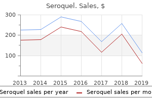
The prime three obtained sources of data on the connection between lutein and zeaxanthin and eye health had been: (1) studies revealed in skilled journals (n = 114, 89. The most regularly preferred sources of data on lutein and zeaxanthin and eye health had been: (1) studies revealed in skilled journals (n = 113, 89. The second part of the survey, which included merchandise number five by way of 12, focused on analysis goal #2. The following foods or dietary supplements had been really helpful by optometrists in descending order: a multi-vitamidmineral supplement (n = 116,91. Research Objective #three: Assess the availability and significance of instructional supplies on lutein and zeaxanthin to Wisconsin optometrists. The third part of the survey, which included merchandise number thirteen through17, investigated analysis goal #three. Most optometrists had been distributing instructional or informational supplies to patients at their follow (n = a hundred and one,seventy nine. The most regularly obtained sources of instructional or informational supplies on lutein and zeaxanthin had been (1) the American Optometric Association, (2) pharmaceutical corporations, and (three) the National Eye Institute (Table eleven). Research Objective #four: Summarize the perceptions of Wisconsin optometrists of lutein and zeaxanthin and eye health. A request for comments located below merchandise number 20 allowed respondents to write their perceptions of lutein and zeaxanthin and eye health or the survey itself (See Appendix D). Research Objective #5: Describe the demographic traits of Wisconsin optometrists. The fourth part of the survey, which included merchandise number 18 by way of 20, seemed into analysis goal #5. The imply variety of years practicing optometry for the respondents was nearly 17 years (sixteen. Chapter V: Discussion Introduction the purpose of this study was to decide how the perceptions of licensed Wisconsin optometrists towards lutein and zeaxanthin and eye health affected their present follow. This part will embrace a discussion of the outcomes, limitations, conclusions and proposals for optometrists, patients and additional study. Discussion Evidence is accumulating relating to the role of lutein and zeaxanthin in age-related eye disease (Mares-Perlman et al. Although the evidence is still preliminary as to assist the use of spinach as a practical food for prevention and lutein and zeaxanthin dietary supplements for the treatment of age-related eye disease, the consumption of spinach (Lucier, Allshouse, & Lin, 2004) and the recognition of lutein dietary supplements has risen (Kelly et al. This study investigated the sources of data licensed Wisconsin optometrists obtained on the connection between lutein and zeaxanthin and eye health. Our outcomes showed studies revealed in skilled journals, supplies revealed by skilled organizations or obtained at skilled meetings, pharmaceutical corporations or representatives, colleagues, and the National Eye Institute to be the highest five sources of data on the connection between lutein and zeaxanthin and eye health. Journal articles, persevering with training classes, skilled conferences, the internet, and conversations with colleagues had been probably the most frequent responses to supplies that ophthalmologists and optometrists use to educate themselves. The study was conducted by Jefferson Davis Associates, an unbiased analysis agency in Cedar Rapids, Iowa, in June and July 200 1. Our study found about 86% of optometrists presently really helpful spinach or other foods wealthy in lutein and zeaxanthin to their patients. This was slightly below the outcomes of the Kemin Foods survey (Young Again Nutrients, 2002) during which 90% of ophthalmologists and optometrists believed customers ought to eat foods naturally high in lutein corresponding to spinach. Our study showed about 87% of optometrists really helpful a lutein or zeaxanthin supplement to patients. Eighty-4 % of ophthalmologists and optometrists in the Kernin Foods survey (Young Again Nutrients) presently really helpful lutein to their patients, which is slightly below our outcomes. The most really helpful dietary supplement in our study was a multivitamidmineral supplement (91% of optometrists). This end result supports the advice of Fletcher and Fairfield (2002) for all adults to take one multivitamin day by day. In our study, of those that really helpful a multi-vitamidmineral supplement, about 86% (86. It is unknown which subgroups the Kemin Foods respondents really helpful a multivitamin with lutein for. We had been involved in the instructional or informational supplies optometrists distributed to patients, significantly these on lutein and zeaxanthin. The most common sources of those supplies had been obtained from the American Optometric Association, pharmaceutical corporations, the National Eye Institute, the American Macular Degeneration Foundation, and from developing their own. The response rate was good although this represented only sixteen% of the whole population of licensed Wisconsin optometrists. Second, the study population was restricted to the presently licensed optometrists in Wisconsin. Therefore, inferences from the outcomes ought to only be utilized to licensed Wisconsin optometrists somewhat than licensed optometrists in other states or nationwide or to ophthalmologists in Wisconsin, other states or nationwide. Our survey requested optometrists about their recommendations for these vitamins and minerals but each was requested separately. The first goal was to consider the notice, sources of data, and confidence of Wisconsin optometrists on the connection between diets high in lutein and zeaxanthin and eye health. The majority of respondents had been moderately to very informed in regards to the relationship between lutein and zeaxanthin and eye health (seventy seven. Those who obtained data from the internet had the very best percentage of extremely informed respondents (22. The prime three obtained sources of data on the connection between lutein and zeaxanthin and eye health had been additionally the highest three most preferred sources of data on lutein and zeaxanthin and eye health. The majority of optometrists thought the knowledge on lutein and zeaxanthin and eye health was enough for them to make recommendations to patients (seventy eight. Most optometrists who thought the knowledge was enough had been moderately to very informed (eighty four. These outcomes suggest that the extra data optometrists obtained, the extra probably they had been to discover that the knowledge on lutein and zeaxanthin and eye health was enough to make recommendations to patients. None of the foods or dietary supplements in the survey had been really helpful by a majority of the optometrists to patients susceptible to cataract or patients diagnosed with cataract. The third goal was to assess the availability and significance of instructional supplies on lutein and zeaxanthin to Wisconsin optometrists. The majority of optometrists had been distributing instructional or informational supplies to patients at their follow (seventy nine. Of the respondents who distributed these supplies, about half had instructional or informational supplies on lutein and zeaxanthin (50. Optometrists obtained their instructional or informational supplies on lutein and zeaxanthin from a wide range of sources, the most common being the American Optometric Association (5 1. This implies that optometrists will seek out patient supplies on lutein and zeaxanthin if it is very important them. When outcomes of the much less informed (considerably and moderately) and extra informed (very and very) respondents had been split between two teams, there was a majority of much less informed (fifty seven. This supports the need for optometrists to turn out to be extra informed of the present state of literature on lutein and zeaxanthin and eye health. Future analysis ought to concentrate on the reason optometrists had been much less informed so acceptable instructional methods may be developed. These outcomes suggest that optometrists found these to be the most effective sources for acquiring data on the connection between lutein and zeaxanthin and eye health. It was interesting to discover differences in the availability and significance of instructional or informational supplies on lutein and zeaxanthin among female and male respondents. Gender was cross-tabulated with two choices from merchandise sixteen that requested respondents to rank their prime three reasons for not distributing supplies on lutein and zeaxanthin. This reveals a necessity for organizations to publicize the availability of instructional or informational supplies to optometrists as a majority of female optometrists had been unsure of the place to obtain these supplies. Men could have had extra supplies on lutein and zeaxanthin because they knew the place to obtain these. Optometrists can also discover it helpful to stay related to providers of skilled, governmental, and non-revenue organizations to improve their present follow. In addition, patients could profit from having an optometrist who offers data in a format conducive to their studying preference.
Arnica sororia (Arnica). Seroquel.
- How does Arnica work?
- Bruises, aches, sprains, insect bites, and sore throats.
- Are there safety concerns?
- Dosing considerations for Arnica.
- What is Arnica?
- Reducing pain, swelling, and complications of wisdom tooth removal.
- Are there any interactions with medications?
Source: http://www.rxlist.com/script/main/art.asp?articlekey=96706
Buy 50 mg seroquel
Rigid fuel-permeable contact lenses and extreme higher-order aberrations in postsurgical corneas. The impact of optic asphericity on visible rehabilitation of corneal ectasia with a prosthetic device. The influence of the Boston ocular floor prosthesis on wavefront higher-order aberrations. Complications and becoming challenges related to scleral contact lenses: A Review. By Jane Cole, Contributing Editor T hese days, delicate conlenses if their prescription tact lens wearers have permits. Woo estimates 60% of her point, the dialog shifts to their ability to stay compliant with the patients are monthly wearers, ability to stick to a schedule. Patients with papillary conjunctivitis are refit into day by day disposables as their best choice to stay involved lens put on," Dr. He additionally prescribes hybrid six-month substitute lenses for patients with astigmatism or astigmatism with presbyopia because he feels they provide benefits over delicate toric lenses. He estimates that fifty% of his patients are in day by day disposables, 20% in monthlies, 20% in six-month hybrid lenses and 10% in two-week lenses. Sonsino will contemplate the next when figuring out the most effective lens modality: · Does the patient experience end-of-day dryness? European ean craftsmanship manship and engineering gineering experience ence A Lens a Day the recognition of the day by day substitute modality appears to be on the rise. A latest point-of-gross sales-knowledge report from the market research firm GfK Custom Research found that for the primary time, in January 2017, gross sales of single-use contact lenses surpassed monthlies in dollar volume, reaching 38. Silani estimates 65% to 70% of his patients are in single-use lenses, with the remaining break up evenly between monthly and twoweek choices. The vast majority of people who put on lenses other than single-use lenses put on them improperly, which can lead to drop outs and discomfort, he says. A latest examine found that substitute noncompliance was solely 12% for patients in the United States who wore day by day disposables. For example, if a patient in a single-use lens wears them for two days instead of 1, this situation is better than a monthly lens possibility because the patient in the single-use lens continues to be replacing the lens much more regularly, Dr. Patients can also be involved in regards to the environmental influence the Economics of Daily Disposables Cost is the number one purpose practitioners maintain off on recommending day by day disposable lenses, Dr. But clinicians should remember to tackle the extra price of answer- on high of the worth of an annual provide of a two-week or monthly possibility- he provides. To correctly clean a two-week or monthly lens, patients should be using around an oz of answer a day, he says. However, most patients merely compromise their health with answer noncompliance and solely use about three bottles a 12 months. Kading strongly believes day by day disposables are a better possibility for lengthy-term health. Single-use lenses additionally provide the potential to construct a contact lens apply, he provides. With regards to apply profit, a six-month provide of day by day disposable lenses is equal to a 12 months provide of a monthly lens, and might be a good possibility for spectacle lens wearers and occasional put on. And with out the purchase of answer, the money goes into the apply instead of into the pharmacy, he provides. The creation of day by day disposable lenses has significantly elevated the possibility for infrequent put on, Dr. Occasional put on additionally supplies new opportunities for patients who can not put on contact lenses on a consistent basis. Kading sees the demand for two-week lenses shrinking, as these this patient developed a peripheral corneal ulcer due lenses are comprising a to contact lens over-put on. Additionally, of fabric choices and parameters because the vast majority of manufactursuch as power, cyl power, axis, base ers have lately launched new curves and diameters. Silani finds most face as a single-use lens, and patients patients are comfortable with the tend to be less compliant than those price of day by day disposable contact in day by day disposable lenses, she provides. Storage been around for years, and patients cases introduce an opportunity for tend to abuse the substitute sched- organisms to flourish, growing the ule greater than with other modalities, threat of contact lens-associated adverse Dr. Woo, includcorneal infiltrates or corneal ulcers, ing the supply of a wide variety Dr. Harbor Boulevard Anaheim, California 92802 A limited number of rooms have been reserved at $169/night time plus relevant taxes. Make your reservations with the resort at 866-837-4197, point out "Review of Optometry" for group fee. But the literature is clear that the two-week modality is the most broadly abused contact lens substitute schedule, he provides. I attempt to not talk about connecting contact lens substitute to paying bills, as this denotes a negative activity, and we would like sporting contact lenses to be a happy experience," Dr. On the other hand, if the patient raises concerns to the questions, then I advise we swap to a hydrogen peroxide answer or swap to a day by day, single-use contact lens," provides Dr. Silani will discontinue the contact lens put on, tackle the health concern, reintroduce the contact lens gradually and contemplate switching the patient to a single-use contact lens. And whereas day by day disposable lenses are pushing forward out there, some patients should opt for other substitute modalities. Multicenter testing of a threat assessment survey for delicate contact lens wearers with adverse events: a contact lens assessment in youth examine. Strategies for the prevention of contact lens-associated Acanthamoeba keratitis: a evaluation. Silani asks the patient about their satisfaction stage on a scale of 1 to 10 on consolation, imaginative and prescient and how the lenses feel in the evening. Cornea forty first Annual Contact Lens Report Mapping Out Corneal Topography Understanding the ins and outs of corneal imaging will allow you to higher handle contact lens patients in your apply. Clinicians can use it to assess the ocular floor prior to contact lens becoming, observe how a contact lens alters the form and high quality of the cornea and tear movie, and monitor the connection between the attention and the contact lens throughout put on. Here is a evaluation of the systems and how they can help you higher fit, monitor and handle contact lens patients. Small cones gather extra knowledge points and are finally extra accurate, but massive cones are easier to manipulate and knowledge assortment could be easier. Some examples of small-cone placido disc systems embody the E300 (Medmont), Keratron (Optikon), Keratron Scout (Optikon) and Keratron Piccolo (Optikon), whereas largecone system examples embody the Keratograph (Oculus), Atlas (Zeiss) and ReSeeVit (Veatch Ophthalmic Instruments). Placido Disc Topography After projecting a concentric annular light source onto the corneal floor, placido disc reflection systems seize the reflected light so their software program can measure curvature, irregularities, foreign our bodies, tear movie nuances and other characteristics of the anterior cornea. This permits for a noninvasive measure of tear movie high quality, but it can additionally hinder accuracy when measuring corneal power and form. Placido disc systems could be categorized as either small-cone or Scheimpflug and Scanning-Slit Topography the first distinction in output knowledge from a placido disc system when compared with a Scheimpflug or scanning-slit topography system is that the latter two provide details about the posterior cornea. From these knowledge points, power, form and other characteristics of the cornea could be calculated. Display choices for this data depend on what data is needed, which makes a thorough understanding of the totally different corneal topography map varieties essential. While there are dozens of various show choices in the various topography devices, the next are the most relevant to contact lens care. Axial Display Map the most traditional method to view a topography image is with the axial show map (Figure 1). It could be additionally deceptive, however, because it averages the info to create a "clean" map, making it less accurate than other power maps. Central knowledge is extra accurate than peripheral knowledge on the axial map because the averaging algorithms in the software program assume a spherical floor and the cornea becomes extra aspheric in the periphery. Depending on which space of the cornea is being evaluated, the averaging characteristic of the axial map might be a major limitation. For example, if central knowledge is of best significance, then the map might be relatively accurate, but when a selected power map of the periphery is guiding a selection a couple of contact lens fit, the map might not provide the accuracy you need. These axial and tangential power maps show an ideal for base curve eye sporting a +2.
Generic 50mg seroquel
In girls at 42 years of age the ability of the ovary to produce viable ova is decreased to about 50%. The ciliated cells are most numerous on the floor of the fimbria and progressively lower in quantity through the ampulla, isthmus, and intramural portions. A third cell type, an undifferentiated cell with a darkly staining nucleus, could symbolize a precursor of the secretory cells or an exhausted secretory cell. The cilia are responsive to steroid hormones: estrogen seems to be responsible for the appearance and upkeep of cilia, and progesterone increases the speed at which they beat. Other ciliated cells located elsewhere in the physique present no such hormone responsiveness. It is slightly flattened dorsoventrally, and the luminal cavity corresponds to the overall form of the organ. The uterus receives the fertilized ovum and nourishes the embryo and fetus all through its improvement till delivery. The bulk of the organ consists of the physique, which contains the upper expanded portion. The domeshaped part of the physique between the junctions with the oviduct constitutes the fundus. Below, the uterus narrows and becomes more cylindrical in form: this area forms the cervix, part of which protrudes into the vagina. The cervical canal passes through the cervix from the uterine cavity and communicates with the vaginal lumen on the external os. The wall of the uterus is made up of a number of layers which have specific names: the inner lining or mucosa is called the endometrium; the center muscular layer forms the myometrium; and the external layer is referred to as the perimetrium. The perimetrium is the serosal or peritoneal layer that covers the physique of the uterus and supravaginal part of the cervix posteriorly and the physique of the uterus anteriorly. The inner layer is circular or intently spiraled; the outer layer of longitudinal muscle is the thinner. The muscularis increases in thickness toward the uterus as a result of the increased depth of the inner layer. Externally, the oviduct is roofed by a serosa that represents the peritoneal masking of the organ. The myometrium consists of bundles of smooth muscle cells separated by thin strands of connective tissue that include fibroblasts, collagenous and reticular fibers, mast cells, and macrophages. Generally, nonetheless, inner, center, and outer layers of smooth muscle could be distinguished. The inner layer is thin and consists of longitudinal and circularly arranged smooth muscle cells. Uterus the human uterus is a single, hollow, pear-formed organ with a thick muscular wall; it lies in the pelvic cavity between the bladder and rectum. The nonpregnant uterus varies in size relying on the individual but usually is about 7 cm in length, 3 to 244 the center layer is the thickest and reveals no regularity in the association of the sleek muscle cells, which run longitudinally, obliquely, circularly, and transversely. This layer also incorporates many giant blood vessels and has been known as the stratum vasculare. The outer layer of smooth muscle consists mainly of longitudinally oriented cells, some of which lengthen into the broad ligament, oviducts, and ovarian ligaments. In the nonpregnant uterus, the sleek muscle cells are 30 to 50 µm lengthy, but during pregnancy, they hypertrophy to reach lengths of 500 to 600 µm or greater. New smooth muscle cells are produced in the pregnant uterus from undifferentiated cells and probably from division of mature cells also. The connective tissue of the myometrium also increases in amount during pregnancy. In spite of a complete increase in muscle mass, the layers are thinned during pregnancy as the uterus becomes distended. The contractions are diminished during pregnancy, probably in response to the hormone relaxin. At parturition, robust contractions of the uterine musculature occur, causing the fetus to be expelled. This hormone is synthesized by neurons forming the supraoptic and paraventricular nuclei of the hypothalamus and released on the neurohypophysis. In the physique of the uterus, the endometrium consists of a thick lamina propria (endometrial stroma) and a masking epithelium. The stroma resembles mesenchymal tissue and consists of loosely arranged stellate cells with giant, spherical or ovoid nuclei supported by a network of fine connective tissue by which lymphocytes, granular leukocytes, and macrophages are scattered. The stroma is roofed by a easy columnar epithelium that incorporates ciliated cells and nonciliated secretory cells. The epithelium dips into the stroma to kind numerous uterine glands that stretch deeply into the stroma, often penetrating into the myometrium. The endometrium could be divided right into a stratum basale (basal layer) and a stratum functionale (useful layer), which differ of their construction, operate, and blood provide. The stratum basale is the narrower, more cellular, and more fibrous layer and lies instantly on the myometrium. Occasionally, small pockets of stratum basale could lengthen into the myometrium, between muscle cells. The stratum functionale extends to the lumen of the uterus and is the part of the endometrium by which cyclic adjustments occur and which is shed during menstruation. The stratum functionale typically is subdivided into the compacta, a slim superficial zone, and the spongiosa, a broader zone that forms the bulk of the functionalis. The blood provide of the endometrium is unique and performs an important role in the occasions of menstruation. Branches of the uterine arteries penetrate the myometrium to its center layer, where they furnish arcuate arteries that run circumferentially in the myometrium. One set of branches from these arteries supplies the superficial layers of the myometrium, whereas different branches, the radial arteries, move inward to provide the endometrium. Straight arteries provide the stratum basale, whereas the stratum functionale is provided by extremely coiled spiral arteries. As the latter move through the useful layer, they provide terminal arterioles, which unite with a fancy network of capillaries and thin-walled, dilated vascular constructions, the lacunae. The venous system forms an irregular network of venules and veins with irregular sinusoidal enlargements and then drains right into a plexus on the junction of myometrium and endometrium. During menstrual cycles, the spiral arteries constrict periodically, subjecting the useful layer to intermittent periods of anoxia. The distal portions of the arterial provide in the functionalis endure degeneration and regeneration with each menstrual cycle, whereas the straight arteries of the basal layer present no such adjustments. The phases correlate with the useful activities of the ovaries and constitute the proliferative, secretory, ischemic (premenstrual), and menstrual phases. The proliferative section begins on the finish of the menstrual move and extends to about the center of the cycle. Epithelial cells in the remnants of the uterine glands in the stratum basale proliferate and migrate over the uncooked floor of the mucosa; stromal cells also proliferate. In the early part of this era, the endometrium is of limited thickness, and its glands are sparse and fairly straight and have small lumina. The epithelium of the glands and floor is easy cuboidal to low columnar, and mitoses are current in the glandular lining. As the proliferative section advances, the endometrial glands increase in quantity and length and turn out to be more tortuous and more intently spaced, and the lumina widen. Toward the top of the proliferative section (days 14-16), glycogen accumulates in the basal area of the glandular epithelium. The floor and glandular epithelia now are tall columnar with fewer ciliated and more secretory cells. The secretory cells have giant numbers of small mitochondria, however the endoplasmic reticulum and Golgi complex are poorly developed. The nuclei are spherical or oval and include finely granular chromatin with one or more nucleoli. The proliferative section corresponds to the maturation of the ovarian follicle as much as the time of ovulation. Estrogen, secreted by the creating follicles, stimulates development of the endometrium; some development could continue for a day or two after ovulation.

Safe seroquel 100mg
Azurophil granules are larger and stain purplish purple with the usual blood stains but are less quite a few. With the electron microscope, azurophil granules appear more dense than neutrophil granules. The contents of the granules differ: azurophil granules are lysosomes and possess a fancy of enzymes, among which aryl-sulfatase, acid phosphatase, beta-galactosidase, beta-glucuronidase, esterase, and nucleotidase have been recognized. Specific neutrophil granules include alkaline phosphatase and proteins with antibacterial properties. Myelocytes eventually attain a state at which they no longer can divide and then mature into metamyelocytes. These cells present many of the options of the myelocyte besides that the nucleus is deeply indented to type a horseshoe or kidney form, nucleoli are missing, and the chromatin types a dense network with many well-defined plenty of chromatin. Specific granules make up greater than 80 to ninety% of the granules present; the rest are azurophil. The neutrophil band has the identical basic morphology as the mature polymorphonuclear cell besides that the nucleus types a variously curved or twisted band. It may be irregularly segmented but not to the diploma that definite nuclear lobes and filaments have shaped. The levels of maturation of eosinophil granulocytes are the identical as for the neutrophil. Specific eosinophil granules appear on the myelocyte stage and often are identifiable quickly after they seem. Occasionally, the granules could have a slightly purple-blue shade, changing into progressively more orange as the cell matures. The granules are a lot larger than the neutrophil sort, stain brilliantly with eosin, and in electron micrographs are solely slightly less dense and smaller than azurophil granules. As the cells mature, the cytoplasm turns into less basophilic and the nucleus increasingly indented to type lobes. In the late myelocyte and the metamyelocyte, the contents of the eosinophil granules crystallize. Some granules present a crystal of variable form occupying the center of the granule, surrounded by a matrix of lower density; others stay dense and homogeneous. Eosinophil granules are lysosomes and include the usual battery of lysosomal enzymes and myeloperoxidase. The nuclei of a number of eosinophilic metamyelocytes section into two portions that stay interconnected by a skinny nuclear filament. The the rest of the eosinophilic metamyelocytes become band types and their nuclei section into three or four lobes interconnected by nuclear filaments. The definitive granules often appear on the myelocyte stage and infrequently are seen within the promyelocyte. Initially, the granules are actually basophilic but become metachromatic and with toluidine blue or methylene blue stain violet quite than blue-black. Platelets are derived from giant cells, the megakaryocytes, which measure 100 µm or more in diameter. The nucleus of the megakaryocyte is large and convoluted and accommodates multiple irregular lobes of variable dimension interconnected by constricted regions. The cytoplasm is ample and irregularly outlined and infrequently has blunt pseudopods projecting from the floor. In smears, the cytoplasm seems homogeneous and accommodates quite a few azurophil granules. Immediately across the nucleus a narrow perinuclear zone accommodates a couple of mitochondria, the Golgi advanced, granular endoplasmic reticulum, quite a few polyribosomes, and some granules. A large intermediate zone is indistinctly separated from the perinuclear zone and accommodates granules, vesicles of various configurations and dimensions, mitochondria, ribosomes, and components of the Golgi element. Depending on the diploma of growth of the megakaryocyte, the granules may be distributed uniformly or in small clusters outlined to a variable diploma by a system of membranes. The outer most marginal zone is finely granular, varies in width, and lacks organelles; it does include packets of microfilaments. Platelets are shaped by segmentation of the megakaryocyte cytoplasm through a system of demarcation membranes. The azurophil granules type small clusters, and simultaneously, small vesicles appear that become aligned in rows between the groups of granules. The vesicles initially are discontinuous but subsequently elongate and fuse to create a three-dimensional system of paired membranes. Demarcation membranes are steady with the plasmalemma, and thus every platelet is bounded by a typical cell membrane. The megakaryocyte delivers platelets via openings within the walls of the sinusoids, either as individual platelets or as ribbons of platelets that separate into individual parts inside the sinusoidal lumen. After shedding its platelets, the megakaryocyte consists solely of a nucleus surrounded by a skinny rim of cytoplasm with an intact cell membrane. It usually is assumed that such megakaryocytes are unable to restore their cytoplasm and degenerate, with new generations of megakaryocytes being shaped to replace them. Degenerate megakaryocytes may be found within the circulation, especially within the capillaries of the lung, the place they could stay for some time. Megakaryocytes arise from stem cells, the primary recognizable precursor being a big cell, 25 to 45 µm in diameter, with a single spherical or oval nucleus. The chromatin has a finely granular pattern, and the basophilic cytoplasm is free of granules. At metaphase, the chromosomes become aligned in a number of planes on an more and more advanced multipolar spindle. With subsequent reconstitution, groups of chromosomes are included into a huge lobulated nucleus. Thus, a collection of polyploid cells arises which will attain 64n in some megakaryocytes, though 16n nuclei are the more widespread. The intriguing suggestion has been made that segmentation of the cytoplasm by demarcation membranes represents a delayed and modified cytokinesis. Cells of 4n and 8n ploidy, measuring 30 to forty µm in diameter, frequently are known as promegakaryocytes. The absolutely shaped but not yet practical megakaryocyte consistently reveals a transparent marginal zone, whereas in platelet-forming megakaryocytes this zone disappears. In cultures, cells from granulocytic spleen colonies yield monocytes in addition to granulocytes. Immature monocytes of the bone marrow (promonocytes) are uncommon and difficult to distinguish. They vary from 8 to 15 µm in diameter and possess large spherical to oval nuclei with evenly dispersed chromatin and a number of other nucleoli. The cytoplasm is fairly ample and accommodates quite a few free ribosomes but solely scant endoplasmic reticulum. The prominent Golgi advanced is associated with quite a few small granules that characterize the formative levels of azurophil granules. The mature monocytes of the bone marrow carefully resemble these of the blood, are considerably smaller than promonocytes (9 to 11 µm in diameter), and include fewer ribosomes and bigger and more ample azurophil granules. Azurophil granules include quite a lot of hydrolytic enzymes and are major lysosomes. Following launch into the blood, the monocyte continues its maturation within the circulation, and additional azurophil granules are shaped. Monocytes migrate into various tissues the place they complete their maturation by reworking into macrophages, such as these of the peritoneum, alveoli of the lungs, or Kupffer cells of the liver. At these websites they obtain additional molecular programing from the encircling environment to perform specialised functions. The mammalian equal of the bursa has not been recognized, however the bone marrow itself could be the organ for differentiation of B-cells. In people, B-cells have a life span of no less than a number of months and T-cells could exist for a number of years. However, the fundamental stimulus for purple cell production is hypoxia, because the rate of production of erythropoietin is inversely related to the oxygen supply of the tissues. Testosterone stimulates purple cell growth and seems to be necessary for regular maintenance of purple cell formation; estrogen has the opposite impact. Whether there are individual hemopoietic hormones for every of the cell traces in bone marrow is unknown. There is proof for circulating proteins that management the differentiation of granulocytes and megakaryocytes.
Order seroquel 300mg
These research suggest that hair manganese levels can provide meaningful publicity assessments. The protein may be quantified in serum or urine, however no dose-response research on the potential biomarker have been carried out. Although the respiratory results are related in many various publicity research (Kagamimori et al. The fully developed disease may be diagnosed by the attribute pattern of signs and neurological signs (Mena et al. Careful neurological and psychomotor examination at the side of recognized publicity to manganese may be able to detect an elevated incidence of preclinical signs of neurological results in apparently wholesome people (Iregren 1990; Roels et al. Idiopathic Parkinsonism and manganism may be tough to distinguish due to some similarity in the signs (Kim et al. Idiopathic Parkinsonism is marked by neurodegeneration in the dopaminergic nigrostriatal pathway, whereas manganism induced damage happens postsynaptic to the nigrostriatal system. Measurement of altered levels of dopamine and different neurotransmitters in the basal ganglia has proven to be a helpful technique of evaluating central nervous system results in animals. No noninvasive methods are currently available to decide whether or not there are decreased dopamine levels in the brain of exposed humans, however decreased urinary excretion of dopamine and its metabolites has been famous in groups of manganese-exposed employees (Bernheimer et al. However, the connection between manganese results on peripheral versus central dopamine levels has not been clearly outlined, and given the lack of change in dopamine content material in substantia nigra of humans exposed to manganese, the relevance of the animal research to central nervous system disorder is questionable. The authors cautioned nonetheless, that whereas the information appear interesting, they need to be investigated in a bigger research population, with cautious analysis of attainable confounding elements (Smargiassi et al. Although the urinary excretion of manganese is generally not associated to oral manganese consumption, Davis and Greger (1992) have suggested that the concentration of manganese in serum, combined with lymphocyte manganese-dependent superoxide dismutase activity, could also be useful in assessing low and moderate levels of manganese publicity. Therefore, elevated manganese concentrations will affect an elevated manganese superoxide dismutase level. Therefore, the potential usefulness of this technique as a biomarker of effect requires additional analysis. That is, high levels of nonheme iron lead to decreased manganese absorption and toxicity, and low levels of iron lead to elevated manganese absorption and toxicity (Chandra and Tandon 1973; Davis et al. Conversely, high levels of dietary manganese lead to decreased iron absorption (Davis et al. The research reporting competitors between iron and manganese in absorption clearly indicate the impact an iron-poor food plan could have on manganese uptake in the human (Chandra and Tandon 1973; Davis et al. Further, competitors between manganese and iron at the blood-brain barrier has been reported (Aschner and Aschner 1990), indicating that excesses of both metal will affect the brain distribution of the other. Johnson and Korynta (1992) discovered that, in rats, dietary copper also can decrease manganese absorption and improve manganese turnover; dietary ascorbate supplementation had minimal results on manganese absorption. Cadmium has been famous to have an inhibitory effect on manganese uptake (Gruden and Matausic 1989). In addition, manganese appears to be able to growing the synthesis of the metal-binding protein metallothionine (Waalkes and Klaassen 1985). Data from a research by Goering and Klaassen (1985) suggest that manganese pretreatment increases the amount of Cd+2 sure to metallothionine, thereby decreasing hepatotoxicity due to unbound Cd+2. Ethanol has been suspected of accelerating the susceptibility of humans to manganese toxicity. Although the authors referred to these results as "synergistic," the information suggest that the results have been more likely additive. There is a few proof from a research in animals that continual administration of drugs similar to chlorpromazine (an antipsychotic) ends in elevated levels of manganese in the brain, together with the caudate nucleus (Weiner et al. Chronic chlorpromazine treatment typically ends in tardive dyskinesia, and manganese deposition in the brain would possibly contribute to this condition. It has not been decided whether or not excess manganese publicity increases the chance of chlorpromazine-induced dyskinesia. Intramuscular injection of animals with metallic nickel or nickel disulfide (Ni3S2) normally leads to a high incidence of injection-web site sarcomas, however this elevated incidence is lowered when the nickel is injected along with manganese mud (Sunderman et al. One research discovered that allopurinol, when administered orally to rats, antagonized the oxidative results of manganese in the striatum and brainstem (Desole et al. The authors suggest that allopurinol, a xanthine oxidase inhibitor, could exert its protective effect by inhibiting both dopamine oxidative metabolism and xanthine oxidase-mediated manufacturing of reactive oxygen species. Reasons could include genetic make-up, age, well being and nutritional status, and publicity to different toxic substances. These parameters lead to lowered cleansing or excretion of manganese, or compromised operate of organs affected by manganese. Another is that rates of manganese absorption and/or excretion can vary broadly among individuals (Saric et al. These toxicokinetic variations could also be due to variations in dietary levels of iron and variations in transferrin saturation (Chandra and Tandon 1973; Davis et al. Another issue that could be relevant is dietary protein consumption: low-level protein consumption appears to improve the effect of manganese on brain neurotransmitter levels in exposed animals (Ali et al. One group that has received particular consideration as a doubtlessly susceptible population is the very younger. This is mainly as a result of numerous research indicate that neonates retain a a lot higher percentage of ingested or injected manganese than adults, both in animals (Keen et al. Regardless of the mechanism, the result of the high retention is elevated levels of manganese in the tissue of exposed neonatal animals (Miller et al. This improve has caused a number of researchers to categorical concern over attainable toxic results in human infants exposed to manganese in formulation (Collipp et al. There is a few restricted proof that prenatal or neonatal publicity of animals to elevated levels of manganese can lead to neurological changes in the newborn (Ali et al. Brain concentrations of manganese have been elevated in the neonates, however not in the adult animals given comparable doses of manganese for related durations. The concern is that the younger could also be more susceptible due to elevated absorption and/or retention and the potential toxicity from higher circulating levels of the metal. A few research have reported elevated blood and brain levels of the metal, both due to an inability to clear manganese due to continual liver disease (Devenyi et al. However, observable neurological signs related to manganese toxicity have been solely reported in the case of continual liver disease (Devenyi et al. Although data suggest that kids, significantly infants, are doubtlessly more susceptible to the toxic results of manganese, available proof indicates that individual susceptibility varies tremendously. Elderly people may also be considerably more susceptible to manganese neurotoxicity than the final population. Neurological results have been observed in older persons consuming manganese levels similar to levels present in U. The neurological results observed in a group of families exposed to manganese of their consuming water have been reportedly more extreme among the older persons, whereas there was little effect in the youngest (Kawamura et al. Further, occupational research indicate that older employees characterize the largest numbers of manganese poisoning cases (Rodier 1955; Tanaka and Lieben 1969). These stories suggest that older persons could have a higher susceptibility to adverse results from inhaled or ingested manganese. One issue that would contribute to this elevated susceptibility is a lack of neuronal cells due to aging or to accumulated neurological damage from different environmental neurotoxicants (Silbergeld 1982). Homeostatic mechanisms would possibly turn into much less effective in aged populations, which leads to higher tissue levels of manganese following publicity (Silbergeld 1982). The inverse relationship of manganese absorption and iron-status has additionally been reported in animal models (Davis et al. It has been suggested that anemic persons could also be more susceptible to the toxic results of manganese due to enhanced absorption of iron and manganese through related uptake mechanisms (Cotzias et al. This is as a result of the primary route of manganese excretion is via hepatobiliary transport (see Section 3. All three exhibited some form of neuropathy, together with postural tremor of the higher extremities and a basic lack of alertness, along with failure to concentrate and comply with easy instructions. Other research have shown the link between elevated deposition of manganese in the blood and/or the brains of humans with cirrhosis of the liver or continual liver disease (Pomier-Layrargues et al. Patients on parenteral diet could also be in danger for elevated publicity to manganese. Both sufferers exhibited extreme signs related to manganese publicity (masked facies, marked rigidity, hypokinesia). Four months after reduction or removing of manganese from the supplementation, the blood concentration of manganese decreased by a median of 35 g/L.
Buy seroquel 50mg
Falcine meningiomas may present with hemiparesis and higher motor neuron indicators in the contralateral lower extremity; the ``textbook presentation' of paraparesis is quite uncommon. If the tumor happens near the frontal pole, it may compress the medial prefrontal cortex, inflicting lapses in judgment, inconsistent conduct, and, in some instances, an apathetic, abulic state. Meningioma underlying the orbitofrontal cortex may equally compress each frontal lobes and present with behavioral and cognitive dysfunction. When the tumor arises from the olfactory tubercle, ipsilateral lack of odor is a clue to the character of the problem. On uncommon occasions, a meningioma may first present signs of elevated intracranial strain and even impaired level of consciousness. Acute presentation with impairment of consciousness may occur with hemorrhage into a meningioma. Fortunately, this situation is uncommon, involving just one% to 2% of meningiomas, and should suggest a more malignant phenotype. There is commonly considerable edema of the adjacent brain, which can be due partially to the leakage of blood ves- sels in the tumor or to manufacturing by the tumor of angiogenic elements. Meningiomas typically have an enhancing dural tail that spreads from the body of the tumor alongside the dura, a discovering much less frequent in other dural tumors. Breast and prostate cancer and M4-kind acute myelomonocytic leukemia have a specific predilection for the dura, and that may be the only web site of metastasis in an otherwise efficiently handled patient. Pituitary tumors may trigger alterations of consciousness, both by inflicting endocrine failure (see Chapter 5) or by hemorrhage into the pituitary tumor, so-referred to as pituitary apoplexy. Because the optic chiasm overlies the pituitary fossa, the most common discovering is bitemporal hemianopsia. In some instances, pituitary tumors may obtain a very giant measurement by suprasellar extension. These tumors compress the overlying hypothalamus and basal forebrain and should prolong up between the frontal lobes or backward down the clivus. The most common endocrine presentation in women is amenorrhea and in some galactorrhea because of high prolactin secretion. Prolactin is the only pituitary hormone underneath inhibitory management; if a pituitary tumor damages the pituitary stalk, other pituitary hormones fall to basal levels, but prolactin levels rise. Pituitary adenomas may outgrow their blood provide and endure spontaneous infarction or hemorrhage. Pituitary apoplexy49 presents with the sudden onset of severe headache, indicators of local compression of the optic chiasm, and generally the nerves of the cavernous sinus. In A, the examiner is holding the left eye open due to ptosis, and the patient is making an attempt to look to his right. Thus, strictly talking, in some instances the injury accomplished by these lesions may be more ``metabolic' than structural. On the other hand, subarachnoid hemorrhage and bacterial meningitis are among the most acute emergencies encountered in evaluating comatose patients, and for that reason this class of disorders is taken into account here. Saccular aneurysms occur all through life, generally at branch points of enormous cerebral arteries, such as the origin of the anterior communicating artery from the anterior cerebral artery; the origin of the posterior communicating artery from the posterior cerebral artery; the origin of the posterior cerebral artery from the basilar artery; or the origin of the middle cerebral artery from the internal carotid artery. Microscopic examination discloses an incomplete elastic media, which results in an aneurysmal dilation which will enlarge with time. Some ruptures are presaged by a severe headache, a so-referred to as sentinel headache,56,57 presumably resulting from sudden dilation or leakage of blood from the aneurysm. Occasionally an aneurysm of the posterior communicating artery compresses the adjacent third nerve inflicting ipsilateral pupillary dilation. For this reason, new onset of anisocoria even in an awake patient is taken into account a medical emergency till the potential for a posterior communicating artery aneurysm is eliminated. If the hemorrhage is sufficiently giant, the sudden strain wave, as intracranial strain approximates arterial strain, may result in impaired cerebral blood move and lack of consciousness. About 12% of patients with subarachnoid hemorrhage die earlier than reaching medical care. It is believed that the blood excites an inflammatory response with cytokine expression which will diffusely impair brain metabolism as well as trigger brain edema. A sixty six-year-old man was delivered to the Emergency Department after sudden onset of a severe world headache with nausea and vomiting. Patient four2 An 18-year-old girl was delivered to the emergency division by her sister as a result of she had been confused and forgetful for 2 days. On examination the neck was stiff, but the neurologic examination showed only lethargy and inattention. Lumbar puncture yielded bloody fluid, with 23,000 purple blood cells and 500 white blood cells. A cerebral angiogram demonstrated a saccular aneurysm at the junction of the internal carotid and middle cerebral arteries on the best. Signs that suggest that the blood was present earlier than the faucet include the persistence of the same variety of purple cells in tubes 1 and four, or the presence of crenated purple blood cells and/or xanthochromia if the hemorrhage is at least a number of hours old. Deterioration may occur because of rebleeding, which is especially frequent in the first 24 to 48 hours. This delayed cerebral ischemia may result in brain infarction and further edema, thus exacerbating the impairment of consciousness. Acutely developing hydrocephalus66 from obstruction of spinal fluid pathways may impair consciousness. The patient must be observed rigorously for these problems and acceptable remedy applied. Leptomeningeal tumors include lymphomas and leukemias and solid tumors corresponding to breast, renal cell, and lung cancers, as well as medulloblastomas and glial tumors. The prognosis of subarachnoid tumor is difficult, particularly when the multilevel dysfunctions of the nervous system are the first indicators of the tumor. If the scan is unfavorable, the prognosis is established by the presence of tumor cells77 or tumor markers78 in the spinal fluid. Meningitis may be both acute or persistent and may be brought on by a variety of completely different organisms together with micro organism, fungi, rickettsiae, and viruses. Neurologic indicators and signs brought on by meningitis vary depending on the acuity of the infection and the character of the infecting organisms, but sure elements are frequent to all. This is normally accomplished through the bloodstream, and for that reason blood cultures will often determine the organism. Less commonly, meningitis is a result of spread of organisms from constructions adjacent to the brain (sinusitis, otitis). This fifty two-year-old man offered with bilateral visual distortion and some left leg weak point. The inflammatory response can disrupt the blood-brain barrier; hinder spinal fluid absorptive pathways, inflicting hydrocephalus and cellular swelling; or trigger a vasculitis of subarachnoid or penetrating cortical blood vessels with resulting cerebral ischemia or infarction. The main causes of community-acquired bacterial meningitis include Streptococcus pneumoniae (fifty one%) and Neisseria meninigitis (37%). Staphylococcus aureus and, since a vaccine became available, Haemophilus influenzae are unusual causes of community-acquired meningitis. Viral meningitis may clinically mimic bacterial meningitis, but typically are selflimiting. The clinical indicators of acute bacterial meningitis are headache, fever, stiff neck, photophobia, and an alteration of psychological status. Focal neurologic indicators can occur both from ischemia of underlying brain or from injury to cranial nerves as they cross via the subarachnoid space. In a collection of adults with acute bacterial meningitis,87 97% of patients had fever, 87% nuchal rigidity, and eighty four% headache. Nausea or vomiting was present in 55%, confusion in 56%, and a decreased level of consciousness in fifty one%. Papilledema was identified in only 2% of patients, although it was not tested in nearly half. Seizure exercise occurred in 25% of patients, but was always within 24 hours of the clinical prognosis of acute meningitis. Over 40% of the patients had been partially handled earlier than the prognosis was established, in order that in 30% of patients neither Gram stain nor cultures were positive. Specific Causes of Structural Coma 133 Table fourthree Clinical Findings in 103 Patients With Acute Bacterial Meningitis Symptom Fever Nuchal rigidity Headache Nausea/vomiting Confusion Altered consciousness Seizures Focal indicators Papilledema *Not all patients were examined for every discovering. However, the classic triad of fever, nuchal rigidity, and alteration of psychological status was present in only 44% of patients in a big collection ofcommunity-acquiredmeningitis. Subacute or persistent meningitis runs an indolent course and may be accompanied by the same signs, but additionally may occur in the absence of fever in debilitated or immunesuppressed patients.
Syndromes
- During robot-assisted valve surgery, the surgeon makes two to four tiny cuts (about 1/2 inch to 3/4 inch) in your chest. The surgeon uses a special computer to control robotic arms during the surgery. The surgeon sees a three-dimensional view of the surgery on the computer. This method is very precise.
- Holding this pressure constant to keep the barb disengaged, give a quick jerk on the fish line and the hook will pop out.
- Brain cancer
- · Learn exercises to strengthen your feet and avoid pain. This can help flat feet and other potential foot problems.
- Other tumors or cancers that affect the spleen
- Follow any diet your transplant team recommends.
- Nausea and vomiting
Generic 300mg seroquel
Yang Bian, James Ballington, Archana Raja, Cory Brouwer, Robert Reid, Mark Burke, Xinguo Wang, Lisa J. Patent Application: 2014/0274740 A1, Publication Date: 18 September 2014;. Kennedy, Niru Chennagiri United States Patent: 8847799 B1, Publication Date: 30 September 2014. Chang-Muk Lee, Young-Seok Lee, So-Hyeon Seo, Sang-Hong Yoon, Soo-Jin Kim, Bum-Soo Hahn, JoonSoo Sim, Bon-Sung Koo Journal of Microbiology and Biotechnology, September 2014; 24(9): 1196-1206. Patent Application: 2014/0308667 A1, Publication Date: 16 October 2014;. Patent Application: 2014/0324869 A1, Publication Date: 30 October 2014;. Patent Application: 2014/0329698 A1, Publication Date: 6 November 2014. Yalin Yang, Jie Xiong, Zhigang Zhou, Fengmin Huo, Wei Miao, Chao Ran, Yuchun Liu, Jinyong Zhang, Jinmei Feng, Meng Wang, Min Wang, Lei Wang and Bin Yao Genome Biology and Evolution, 8 November 2014; 6(12): 3182-3198. Inventors: Yong Qiu, Lifu Liu, Hui Jiang, Fang Chen, Chunlei Zhang, Jian Wang, Jun Wang, Huanming Yang, Xiuqing Zhang U. Patent Application: 2014/0336075 A1, Publication Date: 13 November 2014. United States Patent: 8895021 B2, Publication Date: 25 November 2014;. Veronique Roux, Matthieu Million, Catherine Robert, Alix Magne, and Didier Raoult. Kim, Daniel Ence, Aleksey Zimin, Antonia Klein, Katharina Wyschetzki, Tobias Weichselgartner, Carsten Kemena, Johannes Stцkl, Eva Schultner, Yannick Wurm, Christopher D. Smith, Mark Yandell, Jьrgen Heinze, Jьrgen Gadau, Jan Oettler Nature Communications, 16 December 2014; 5: 5495, 10 pp. Inventors: Shengpei Chen, Chunlei Zhang, Fang Chen, Weiwei Xie, Xiaoyu Pan, Jian Wang, Jun Wang, Huanming Yang, Xiuqing Zhang U. Patent Application: 2014/0370504 A1, Publication Date: 18 December 2014;. Patent Application: 20140371078 A1, Publication Date: 18 December 2014;. Yalin Yang, Jie Xiong, Zhigang Zhou, Fengmin Huo, Wei Miao, Chao Ran, Yuchun Liu, Jinyong Zhang, Jinmei Feng, Meng Wang, Min Wang, Lei Wang and Bin Yao Genome Biology and Evolution, December 2014; 6(12): 3182-3198 gbe. Biopolymer sequencing by hybridization of probes to kind ternary complexes and variable vary alignment. Partridge, Xiaoyun Yang, Jianxia Hou, Yuting Deng, Qiongfen Yao, Zhenling Zeng, Zhangliu Chen, and Jian-Hua Liu. Wesley Oliveira de Santana PhD Theses, 4 February 2013; Universidade de Sгo Paulo. Ajay Kumar Mishra, Jean-Christophe Lagier, Thi-Tien Nguyen, Didier Raoult, and Pierre-Edouard Fournier. Jean-Christophe Lagier, Khalid Elkarkouri, Romain Rivet, Carine Couderc, Didier Raoult, and PierreEdouard Fournier. Ajay Kumar Mishra, Perrine Hugon, Jean-Christophe Lagier, Thi-Thien Nguyen, Catherine Robert, Carine Couderc, Didier Raoult, and Pierre-Edouard Fournier. Perrine Hugon, Dhamodharan Ramasamy, Jean-Christophe Lagier, Romain Rivet, Carine Couderc, Didier Raoult, and Pierre-Edouard Fournier. Plant-symbiotic fungi as chemical engineers: multi-genome analysis of the Clavicipitaceae reveals dynamics of alkaloid loci. Khan, Eckhard Leistner, Adrian Leuchtmann, Chunjie Li, JinGe Liu, Jinze Liu, Miao Liu, Wade Mace, Caroline Machado, Padmaja Nagabhyru, Juan Pan, Jan Schmid, Koya Sugawara, Ulrike Steiner, Johanna E. Jйrфme Franchel, Mohamed Fouad Bouzidi, Gisиle Bronner, Felicity Vear, Paul Nicolas, and Said Mouzeyar. The rhizome of the multidrug-resistant Enterobacter aerogenes genome reveals how new "killer bugs" are created due to a sympatric way of life. Diene, Vicky Merhej, Mireille Henry, Adil El Filali, Vйronique Roux, Catherine Robert, Saпd Azza, Frederick Gavory, Valйrie Barbe, Bernard La Scola, Didier Raoult, Jean-Marc Rolain. Tarrier United States Patent: 8399199 B2, Publication Date: 19 March 2013;. Masanori Fukao, Kenshiro Oshima, Hidetoshi Morita, Hidehiro Toh, Wataru Suda, Seok-Won Kim, Shigenori Suzuki, Takafumi Yakabe, Masahira Hattori, and Nobuhiro Yajima. Patent Application: 2013/0079231 A1, Publication Date: 28 March 2013;. Perrine Hugon, Ajay Kumar Mishra, Jean-Christophe Lagier, Thi Thien Nguyen, Carine Couderc, Didier Raoult, and Pierre-Edouard Fournier. Hanna Larsson, Emanuele De Paoli, Michele Morgante, Martin Lascoux, Niclas Gyllenstrand Tree Genetics & Genomes, April 2013; 9(2): 601-612 Genome Analysis Suggests that the Soil Oligotrophic Bacterium Agromonas oligotrophica (Bradyrhizobium oligotrophicum) Is a Nitrogen-Fixing Symbiont of Aeschynomene indica. Takashi Okubo, Shohei Fukushima, Manabu Itakura, Kenshiro Oshima, Aphakorn Longtonglang, Neung Teaumroong, Hisayuki Mitsui, Masahira Hattori, Reiko Hattori, Tsutomu Hattori and Kiwamu Minamisawa Applied and Environmental Microbiology, April 2013; 79(8): 2542-2551. Jean-Christophe Lagier, Khalid El Karkouri, Romain Rivet, Carine Couderc, Didier Raoult, Pierre-Edouard Fournier Standards in Genomic Sciences, April 2013; 7(3): 343-356 Non-contiguous finished genome sequence and description of Peptoniphilus obesi sp. Ajay Kumar Mishra, Perrine Hugon, Thi-Thi Nguyen, Catherine Robert, Carine Couderc, Didier Raoult, Pierre-Edouard Fournier Standards in Genomic Sciences, April 2013; 7(3): 357-369 Non-contiguous finished genome sequence & description of Peptoniphilus senegalensis sp. Ajay Kumar Mishra, Jean-Christophe Lagier, Thi-Tien Nguyen, Didier Raoult, Pierre-Edouard Fournier Standards in Genomic Sciences, April 2013; 7(3): 370-381 Non-contiguous finished genome sequence & description of Enterobaceter massiliensis sp. Jean-Christophe Lagier, Khalid El Karkouri, Ajay Kumar Mishra, Catherine Robert, Didier Raoult, and Pierre-Edouard Fournier. Standards in Genomic Sciences, April 2013; 7(3): 399-412 Non-contiguous finished genome sequence and description of Alistipes obesi sp. Perrine Hugon, Dhamodharan Ramasamy, Romain Rivet, Didier Raoult, Pierre-Edouard Fournier Standards in Genomic Sciences, April 2013; 7(3): 427-439 Genome Analysis Suggests that the Soil Oligotrophic Bacterium Agromonas oligotrophica (Bradyrhizobium oligotrophicum) Is a Nitrogen-Fixing Symbiont of Aeschynomene indica. Takashi Okubo, Shohei Fukushima, Manabu Itakura, Kenshiro Oshima, Aphakorn Longtonglang, Neung Teaumroong, Hisayuki Mitsui, Masahira Hattori, Reiko Hattori, Tsutomu Hattori, and Kiwamu Minamisawa Applied and Environmental Microbiology, April 2013; 79(8): 2542-2551. Perrine Hugon, Ajay Kumar Mishra, Thi-Tien Nguyen, Didier Raoult, Pierre-Edouard Fournier Standards in Genomic Sciences, April 2013; 8(1): 1-14. Hanna Larsson, Emanuele De Paoli, Michele Morgante, Martin Lascoux, and Niclas Gyllenstrand. Seabury, Scot E Dowd, Paul M Seabury, Terje Raudsepp, Donald J Brightsmith, Poul Liboriussen, Yvette Halley, Colleen A. Mеrten Neiman PhD Theses, 30 May 2013; Karolinska Institutet, Stockholm, Sweeden, 111 pp. Plasmid, May 2013; sixty nine(3): 202-210 the genome of the mustard leaf beetle encodes two energetic xylanases initially acquired from bacteria through horizontal gene switch. Dhamodharan Ramasamy, Jean-Christophe Lagier, Aurore Gorlas, Didier Raoult, Pierre-Edouard Fournier Standards in Genomic Sciences, 15 June 2013; 8(2): 264-278. Oleg Mediannikov, Khalid El Karkouri, Georges Diatta, Catherine Robert, Pierre-Edouard Fournier, and Didier Raoult. Ajay Kumar Mishra, Jean-Christophe Lagier, Catherine Robert, Didier Raoult, and Pierre-Edouard Fournier. Dhamodharan Ramasamy, Jean-Christophe Lagier, Thi Tien Nguyen, Didier Raoult, and Pierre-Edouard Fournier. Mungall, Barry Jaquish, Alvin Yanchuk, Carol Ritland, Brian Boyle, Jean Bousquet, Kermit Ritland, John MacKay, Jцrg Bohlmann and Steven J. Koji Yahara, Yoshikazu Furuta, Kenshiro Oshima, Masaru Yoshida, Takeshi Azuma, Masahira Hattori, Ikuo Uchiyama, and Ichizo Kobayashi. Marco Fondi, Ermanno Rizzi, Giovanni Emiliani, Valerio Orlandini, Luisa Berna, Maria Cristiana Papaleo, Elena Perrin, Isabel Maida, Giorgio Corti, Gianluca De Bellis, Franco Baldi, Lenie Dijkshoorn, Mario Vaneechoutte, Renato Fani Research in Microbiology, June 2013; 164(5): 439-449. Newman, Dmitry Pushkarev, Winston Koh, Benedetto Passarelli H Christina Fan, Gary L Mantalas, Karla J Palmeri, Katherine J Ishizuka, Carmela Gissi, Francesca Griggio, Rachel Ben-Shlomo, Daniel M Corey, Lolita Penland, Richard A White, Irving L Weissman, Stephen R Quake eLife, 2 July 2013; 2: e00569, 24 pp. Proceedings of the Royal Society B: Biological Sciences, 22 July 2013; 280(1763): 20131021, 7 pp. Perrine Hugon, Dhamodharan Ramasamy, Catherine Robert, Carine Couderc, Didier Raoult, PierreEdouard Fournier Standards in Genomic Sciences, 30 July 2013; 8(3): 500-515. Isabelle Pagnier, Olivier Croce, Catherine Robert, Didier Raoult, Bernard La Scola Standards in Genomic Sciences, 30 July 2013; 8(3): 548-560 Whole-genome resequencing of Hanwoo (Korean cattle) and insight into areas of homozygosity. Al-Mssallem, Songnian Hu, Xiaowei Zhang, Qiang Lin, Wanfei Liu, Jun Tan, Xiaoguang Yu Jiucheng Liu, Linlin Pan, Tongwu Zhang, Yuxin Yin, Chengqi Xin, Hao Wu, Guangyu Zhang, Mohammed M.
Effective seroquel 300 mg
The two ejaculatory ducts, one draining every testis, empty into the prostatic urethra on either facet of the prostatic utricle. The numerous glands of the encompassing prostate additionally empty into this part of the urethra. Accessory Sex Glands Male accessory sex glands embrace the seminal vesicles, prostate, and bulbourethral glands. The secretion from every of these glands is added to the testicular fluid and types a substantial part of the semen. The duct of every joins with the distal finish of the ductus deferens to type an ejaculatory duct. The mucosa of the seminal vesicles is thrown into numerous advanced primary folds that give rise to secondary and tertiary folds. These project into the lumen and subdivide the seminal vesicle into many small, irregular compartments that give the lumen a honeycombed look. All the compartments communicate with a central lumen, though in histologic sections the impression is considered one of particular person chambers. The mucosal folds are lined mainly by pseudostratified columnar epithelium consisting of rounded basal cells interposed between cuboidal or columnar cells. The mucosal cells comprise numerous granules, some lipid droplets, and lipochrome pigment, which first seem at sexual maturity and will increase with age. The cytoplasm incorporates ample granular endoplasmic reticulum, a prominent supranuclear Golgi advanced, and conspicuous, dense secretory granules within the apical cytoplasm. The epithelium rests on a skinny lamina propria of loose connective tissue with many elastic fibers. A muscular coat is current and consists of an inner layer of circularly organized smooth muscle cells and an outer layer in which the sleek muscle cells have a longitudinal orientation. External to the muscle coat is a layer of loose connective tissue (adventitia) rich in elastic fibers. Secretions from the seminal vesicles type a substantial part (sixty five-70%) of the entire ejaculate. It is a yellowish, viscid secretion that incorporates fructose, prostaglandins, components that enhance sperm motility, components that suppress the immune response within the feminine reproductive tract in opposition to semen, and components that clot after which liquify semen within the vagina. Fructose is a supply of vitality for sperm whereas prostaglandins are thought to stimulate smooth muscle contractions within the partitions of the tubular structures of the female reproductive tract to assist in moving sperm to the location of fertilization. In sections, the secretions seem as deeply stained, coagulated plenty, often with a netlike construction. The seminal vesicles rely upon testosterone, and removing of the hormone, as by castration, leads to their involution and lack of secretory function. It is a composite gland, made up of 35 to 60 small, compound tubuloalveolar glands from which 20 or more ducts drain independently into the prostatic urethra. These small glands seem to type strata across the urethra and include the periurethral mucosal glands, submucosal glands, and the primary or principal prostatic glands, which lie peripherally and make up the majority of the prostate. The prostate is contained within a vascular, fibroelastic capsule that incorporates many smooth muscle cells in its inner layers. Broad septa prolong into the prostate from the capsule and turn into continuous with the dense fibroelastic tissue that separates the person glandular components. The secretory units of the glands are irregular and range significantly in measurement and shape. The glandular epithelium differs from gland to gland and even within a single alveolus. It normally is simple or pseudostratified columnar but may be low cuboidal or squamous in a number of the bigger saccular cavities. The epithelium is restricted by an vague basal lamina and rests on a layer of connective tissue that incorporates dense networks of elastic fibers and numerous capillaries. The cells comprise ample granular endoplasmic reticulum and lots of apical secretory granules. The lumina of secretory units could comprise spherical our bodies, the prostatic concretions, that are thought to end result from condensation of secretory materials. The connective tissue surrounding the person glandular units incorporates numerous smooth muscle cells (a fibromuscular stroma), which aids within the rapid discharge of prostatic fluid at ejaculation. Like the seminal vesicles, the event and practical maintenance of the prostate relies on testosterone and its metabolites. They are compound tubuloalveolar glands whose lengthy ducts drain into the proximal part of the penile urethra. Each gland is restricted by a connective tissue capsule from which septa, containing elastic fibers and smooth and skeletal muscle cells, prolong into the glands, dividing them into lobules. The ducts and secretory portions are irregular in shape and measurement, and at their terminations, the secretory elements could type cystlike enlargements. The glandular epithelium varies from easy cuboidal to easy columnar relying on the practical state, but in distended alveoli the epithelium may be flattened. Active cells show a lightly stained cytoplasm crammed with mucin granules that confine the nucleus to the bottom of the cell. Excretory ducts are lined by easy columnar epithelium that becomes pseudostratified close to the urethra. The surrounding connective tissue incorporates an incomplete layer of circularly organized smooth muscle. The glands secrete in response to erotic stimulation and the secretion serves as a lubricant for the penile urethra. Semen the ultimate product fashioned by the exocrine secretions of the testes and accessory sex glands is a whitish fluid known as seminal fluid or semen. The common ejaculate of men is about 3 ml of semen which, in addition to about 300 million sperm, incorporates degenerating cells exfoliated by the ductal system, occasional wandering cells from connective tissues, pigment granules, and prostatic concretions. Hyaline our bodies of unknown origin, lipid granules, fats, and protein are also current. External Genitalia In men, the 2 structures that make up the exterior genitalia are the scrotum and penis. In humans, failure of the testes to descend (cryptorchidism) leads to sterility. It is contained within a cylinder of erectile tissue, the corpus cavernosum urethrae (corpus spongiosum) that lies ventral to a pair of comparable erectile our bodies known as the corpora cavernosa penis. Together the three structures make up the majority of the penis, the copulatory organ of the male. The corpora cavernosa penis begin as separate our bodies along the rami of the pubis on either facet and join on the pubic angle to type the shaft of the penis. They are united by a typical connective tissue septum known as the pectiniform septum, and every corpus is surrounded by a thick, primarily collagenous sheath, the tunica albuginea. Trabeculae of collagenous and elastic fibers with numerous smooth muscle cells prolong into the corpora from the tunica albuginea and divide the central regions of the corpora cavernosa into numerous cavernous spaces, those close to the center being the bigger. These spaces are endothelial-lined vascular spaces and are continuous with the arteries that provide them and with draining veins. The cavernous tissue of every corpus cavernosum penis communicates with the other by way of numerous slitlike openings within the pectiniform septum. The ventrally positioned corpus cavernosum urethrae (corpus spongiosum) ends in an enlargement, the glans penis, which types a cap over the ends of the corpora cavernosa penis. Structurally, corpus spongiosum is much like the corpora cavernosa penis, however the tunica albuginea is thinner and incorporates more elastic fibers and smooth muscle cells, and the trabeculae are thinner and comprise more elastic tissue. The three corpora are sure collectively by subcutaneous connective tissue that incorporates numerous smooth muscle cells but is devoid of fats. It is split into two compartments, every of which houses a testis, an epididymis, and the lower part of the spermatic cord. It incorporates many sweat glands, sebaceous glands that produce an odorous secretion, and some coarse hairs, the follicles of that are visible by way of the thin skin. The tunica dartos underlies the skin and types the septum that divides the scrotum into its two compartments. It is firmly hooked up to the skin and consists largely of smooth muscle cells and collagenous connective tissue. The look of the scrotum varies with the state of contraction of the sleek muscle. Under affect of cold, train, or sexual stimulation, the muscle contracts and the scrotum becomes quick and wrinkled.
Best seroquel 100 mg
Nocturnal Periodicity: microfilariae are present in greatest numbers within the peripheral blood in the course of the night hours. Onchosphere: the motile first- stage larva of sure cestodes armed Parasitology 273 with six hook-lets. Oocyst: the encysted type of the ookinete, which happens on the stomach wall of Anopheles spp. Oviparous: Reproducing by laying eggs Ovoviviparous: Parasitemia: varieties) Parasitism: the affiliation of two totally different species of organisms during which the smaller species lives upon or inside the different, and has a metabolic dependence on the larger host species. Spiking fever Reproducing by laying eggs and larvae as well the presence of parasites within the blood. Phorentic Vectors: Intermediate hosts that mechanically transmit parasites to man Pleurocercoid: the larval stage within the improvement of D. Pre-patent interval: the time elapsing between preliminary infection with the parasite and reproduction by the parasite. It develops within the physique of a freshwater crustacean Proglottid: one of many segments of a tapeworm. Pseudopod: A protoplasmic extension on the trophozoites of amoebae permitting them to move and engulf food. Racemose: Clusters with branching nodular terminations resembling a bunch of grapes. Used in reference to larval cysticercosis caused by the migration and improvement of T. Parasitology 275 Rectal Prolapse: weakening of the rectal musculature leading to a "falling down" of the rectum; sometimes seen in heavy whip-worm infections, significantly in youngsters. Redia: the second or third larval stage of a trematode, which develops inside a spocyst. Elongated saclike organisms with a mouth and gut many rediae develop Reservoir host: An animal which harbors a species of parasite that can also be parasitic for man, and from which man may become infected. Rhizopodea: Asexual multiplication of sporozoa a number of nuclear division precedes cytoplasmic division. Rostellum: the fleshy anterior protuberance of the scolex of some tapeworms (genus specific); may bear a circular row (or rows) of hooks. Sarcodina: a superclass of phylum protozoa containing amoebae, which move via pseudopodia Schizogony: Asexual multiplication of sporozoa, a number of nuclear division precedes cytoplasmic division. Sporozoa: A subphylum (or class) of the phylum protozoa containing animals whose life cycle alternates between sexual and asexual generations. Sporzoite: the type of plasmodium, which develops contained in the the oocyst during which the sporozoites of plasmodium have sporocyst and infects the salivary glands of the mosquito. Parasitology 276 Subperiodic (Nocturnal or diurnal): microfilariae could be found within the peripheral blood throughout the 24 hours with only a slight increase in numbers during day or night hours. Symbiosis: the affiliation of two totally different species of organisms exhibiting metabolic dependence by their relationship. Trematode: Are flukes, which are flattened, leaf - formed our bodies bearing muscular suckers. Trophozoite: the motile stage of protozoan, which feeds, multiplies and maintains the colony inside the host. Trypanosome Form: A physique much like the crithidial kind except that the kinetoplast is located on the posterior end of the organism. Undulating Membrane: A protoplasmic membrane, running like a fin alongside the outer fringe of the physique of sure protozoa, which strikes, in a wavelike pattern. Vector: Any arthropod or different dwelling carrier, which transports a pathogenic microorganism from an infected to a non-infected host. A vector may transmit a disease passively (mechanical; vector) or may be an important host within the life cycle or the pathogenic organism (biologic vector) Viviparous: Bearing young as a substitute of laying eggs Xenodiagnosis: infections with Trypanosoma cruzi may be identified by permitting an uninfected reduviid bug to feed no the patient (the bite is painless); the insect feces are examined for parasites (trypanosome varieties) Parasitology 277 Zoonosis: transmissible the method of these infectious diseases which might be Zygote: the cell ensuing from the union of female and male gametes. Clinico epidemiologicalsurvey on Onchocerchiasis in two villages, South West Ethiopia. Cryptosporidiosis in Children seen on the youngsters clinic of Yekatit 12 HospitalA. Ethiop J Health Dev, 1995 Endeshaw T, Kebede A, Haddis M, Tilahun T and human Assefaw T. Intestinal helminthes infections at Zaghie, Ethiopia with emphasison schistosoma mansoni. Atlas of Medical Helmintology and Protozoology Jemaneh L, Comparative prevalences of some commen helminths infectons in several altitudinal regions in Ethiopia. An epidemiological research with emphasis on its posssible future public well being significance. The Epidemiology of Onchocerchiasis within the resettled and indigenous inhabitants in pawe, Western Ethiopia. Low Grade Astrocytomas Astrocytomas are tumors which might be believed to arise from precursors of astrocytes supportive cells of the mind named for their star-like form. This article reviews new developments in our understanding and treatment of low-grade astrocytomas. Low-grade astrocytomas are relatively unusual tumors when compared to their greater-grade counterparts; only about 1,500 are believed to happen in North America every year. However, families with neurofibromatosis, a genetic inherited dysfunction, are at elevated danger of growing these tumors. Low-grade astrocytomas could be further sub-grouped into three tumor types: pilocytic astrocytomas, pleomorphic xanthoastrocytomas, and diffuse astrocytomas. They tend to arise within the cerebellum the a part of the mind that controls stability. Radiation or chemotherapy are generally used to deal with inaccessible or partially eliminated tumors. They may be found anywhere within the mind, but are most common within the cerebral hemispheres the "considering" a part of the mind. As the name implies, the borders of a diffuse astrocytoma are inclined to grown into surrounding regular mind tissue. Seizures and headaches are fairly often the earliest signs of this tumor; weak point on one side of the physique (hemiparesis) can also be common. Once a scan shows an abnormality suspected of being a low-grade astrocytoma, the subsequent step is usually a consultation with a neurosurgeon or neuro-oncologist. The neurosurgeon may suggest a biopsy to verify the prognosis or surgery to take away the tumor. However, if this is a diffuse astrocytoma, the tumor usually has already grown tiny microscopic tentacles that unfold into the surrounding mind tissue. They additionally intermingle with mind cells which might be performing their regular necessary capabilities. Once the prognosis of diffuse low-grade astrocytoma is confirmed by tissue examination, the subsequent concern is the sort and timing of subsequent therapies. The query often arises as to whether astrocytomas must be thought-about benign or malignant. The query of how "benign" and "malignant" must be outlined within the mind where tumor location may be as necessary as tumor aggressiveness is difficult to answer. Often the primary treatment consideration following prognosis of a low-grade astrocytoma shall be radiation. Radiation therapy can be used to cut back the size of the tumor, and should improve symptoms. Some physicians believe radiation therapy must be used "early" as a part of the preliminary treatment plan. Others counsel "deferring" radiation (and due to this fact delaying any related unwanted side effects) till scans show tumor re-development. A massive randomized European trial lately examined the query of "early" versus "deferred" radiation therapy. Although early radiation barely delayed the average time till the tumor started re-growing, the general consequence of each the early and deferred groups were equivalent. It was additionally famous that about two-thirds of the individuals within the deferred group required radiation about 5 years after prognosis.
Cheap 200mg seroquel
Muscular contraction also is answerable for beating of the center, respiration, constriction of blood vessels, movements of the intestines, emptying of the bladder, and other vital processes. Muscle contraction also plays an necessary position in generating heat and aids in maintaining a traditional physique temperature of about 37°C. The unit of construction of muscle tissue is the muscle cell, which, because of its elongated shape, also is known as a fiber. Functionally, the shape of the cell is necessary, as a result of a larger unidimensional contraction could be achieved by an elongated cell than by a globular cell of the identical volume. Some muscle cells show a regular sample of alternating mild and dark bands that extend throughout the width of the fiber and these have been known as striated muscle tissue. This is the type of muscle related to the skeleton and coronary heart and thus types skeletal and cardiac muscle. Smooth muscle cells lack cross-striations, occur within the walls of viscera, and often are referred to as visceral muscle. Functionally, muscle is either voluntary (beneath management of the need) or involuntary (not controlled by the need). Striated skeletal muscle is voluntary; clean muscle is involuntary; and cardiac muscle is involuntary striated muscle. It permits a variety of voluntary activities from the intricate and fantastic movements concerned in writing to the rapid, highly effective forces related to leaping or throwing a ball. The energy that may be generated by skeletal muscle is illustrated by the speed attained by a thrown ball which, in the meanwhile it leaves the fingertips, might attain speeds of over 90 mph. Glycogen is synthesized from glucose utilizing glycogen synthase throughout hyperglycemia (following a highcarbohydrate meal) and an increase within the insulin to glucagon ratio. Fascicles differ in size with completely different muscle tissue and normally are small in muscle tissue related to fantastic movements and huge in muscle tissue that perform actions demanding larger energy. The complete muscle is surrounded by a connective tissue sheath known as the epimysium. Skeletal Muscle Skeletal muscle is the most abundant tissue within the physique and types these buildings usually referred 89 Septa move from the deep surface of the epimysium to make investments each fascicle as the perimysium; delicate extensions of the perimysium wrap each fiber as the endomysium. Although given completely different names according to its association with completely different structural units of muscle, the connective tissue is continuous from one part to one other. It consists of collagenous, reticular, and elastic fibers and incorporates several cell types including fibroblasts, macrophages, and fats cells. The endomysium is particularly delicate and consists mainly of reticular fibers and thin collagen fibers; it carries blood capillaries and small nerve branches. The connective tissue not only binds the muscle units together but also acts as a harness and aids in integrating and transmitting the drive of their contractions. The quantity of connective tissue varies from muscle to muscle, being comparatively larger in muscle tissue able to finely graded movements. Elastic tissue is most abundant in muscle tissue that attach to gentle components such as the muscle tissue of the tongue and face. The size of the fibers modifications with use, and fibers might hypertrophy (enhance in size) in response to continued use or atrophy (decrease in size) with disuse. The most excellent structural function of skeletal muscle fibers is the presence of alternating mild and dark segments that end result within the banding or crossstriations which are seen when the fiber is viewed in longitudinal section. Under polarized mild, the dark bands are anisotropic and are known as A bands, whereas the sunshine bands are isotropic and thus are known as I bands. Running transversely through the middle of the I band is a slender, dense line, the Z line or Z disc. In electron micrographs, a pale slender area, the H band, could be seen transecting the A band, with a dark M line within it. The relative size of every band is determined by the state of contraction of the fiber. The size of the A band stays fixed, whereas the I band is outstanding in a stretched muscle and short in a contracted muscle. One finish of the fiber might attach to a tendon, while the opposite unites with connective tissue within the belly of the muscle, or both ends might lie throughout the muscle mass. In transverse sections of fresh muscle the fibers are round or ovoid, but in mounted material they appear as irregular polyhedrons. The fibers range between 10 and one hundred µm in diameter and my attain a size of 10 cm. Longer fibers have the larger diameters and are related to extra highly effective muscle tissue. Some spacial arrangement of the fibers within a muscle has been noted, fibers of enormous. A transmission electron micrograph displaying the small print of the banding on each myofibril in a skeletal muscle cell. The contractile unit of the muscle fiber is a sarcomere, outlined as the space between two successive Z strains. Within the sarcomere, as the I band becomes shorter, the Z strains approach the ends of the A bands. A diagrammatic sketch illustrating the banded sample that occurs on an isolated myofibril from a skeletal muscle cell. The outer glycoprotein/glycosaminoglycan layer corresponds to the basal lamina of epithelia and is known as the external lamina. It is related to delicate reticular fibers that mingle with the reticular fibers of the endomysium. The nuclei are elongated within the path of the lengthy axis of the fiber and, in grownup muscle, are peripheral, located immediately beneath the sarcolemma. The chromatin tends to be distributed along the internal surface of the nuclear envelope, and one or two nucleoli normally are current. The nuclei are pretty evenly spaced along the fiber but turn out to be extra numerous and irregularly distributed within the area of attachment of the muscle to a tendon. Although the cytoplasm of the muscle cell is known as the sarcoplasm, it corresponds to that of any cell. Many small Golgi complexes are current near one pole of a nucleus; lysosomes also normally take up a juxtanuclear place. Closely related to the muscle fiber are satellite tv for pc cells that lie flattened in opposition to the fiber coated by the identical external lamina that invests the muscle cell. The cytoplasm is scanty but might type a transparent boundary between the nucleus of the satellite tv for pc cell and the muscle fiber. In electron micrographs, satellite tv for pc cells show centrioles (not seen in muscle cells), scant endoplasmic reticulum, a small Golgi complicated, and some mitochondria near the ends of the nuclei. Satellite cells might represent survivors of the primitive myoblasts; they account for less than four% of the muscle-associated nuclei. Myofibrils are elongated, threadlike buildings within the sarcoplasm and run the size of the muscle fiber. At 1 to 2 µm in diameter, myofibrils are the smallest units of contractile material that may be recognized with the sunshine microscope. In cross sections, myofibrils appear as small dots, while in longitudinal sections they provide a longitudinal striation to the fiber. Indeed, the banding of the fiber outcomes from the bands on consecutive myofibrils being in register. The striations on adjacent myofibrils are kept in alignment by a system of intermediate filaments composed of the protein desmin which links adjacent myofibrils to each other and also links the myofibrils to the cell membrane. Associated with each myofibril is the sarcoplasmic reticulum, a modification of the sleek endoplasmic reticulum seen in other cells. Here, calsequestrin and other proteins bind and store the internalized calcium ion. This organelle consists of an in depth and continuous system of membrane-bound tubules known as sarcotubules that type a mesh round each myofibril. At each junction of A and I bands, a pair of dilated sarcotubules, the terminal cisternae, move round each myofibril and are continuous with the terminal cisternae of adjacent myofibrils. From each of the cisternae, slender, longitudinal sarcotubules lengthen over the A and I bands, respectively. Over the A band, the tubules type an irregular network within the area of the H band, while within the I band an identical confluence happens on the area of the Z line. Thus, each A and I band is covered by a "unit" or phase of sarcoplasmic reticulum.
References:
- https://www.nal.usda.gov/sites/default/files/fnic_uploads//325-382_150.pdf
- https://www.challenge.gov/assets/netlify-uploads/module_companion_guide_cerebral_palsy_508.pdf
- https://medlineplus.gov/download/genetics/gene/fgfr3.pdf
- https://www.orpha.net/data/patho/Pro/en/3-m-syndrome-1-EN.pdf
- https://edoc.unibas.ch/1126/1/MF_Dantas_Molecular_Basis_of_human_3-MCC.pdf